Atypical Fournier’s presenting with pneumoscrotum, a testicular tumour diagnosed wrongly as hydrocele, a recurred dermatofibrosarcoma and a GIST of ileum
Posted on: October 27, 2019
13.4.19
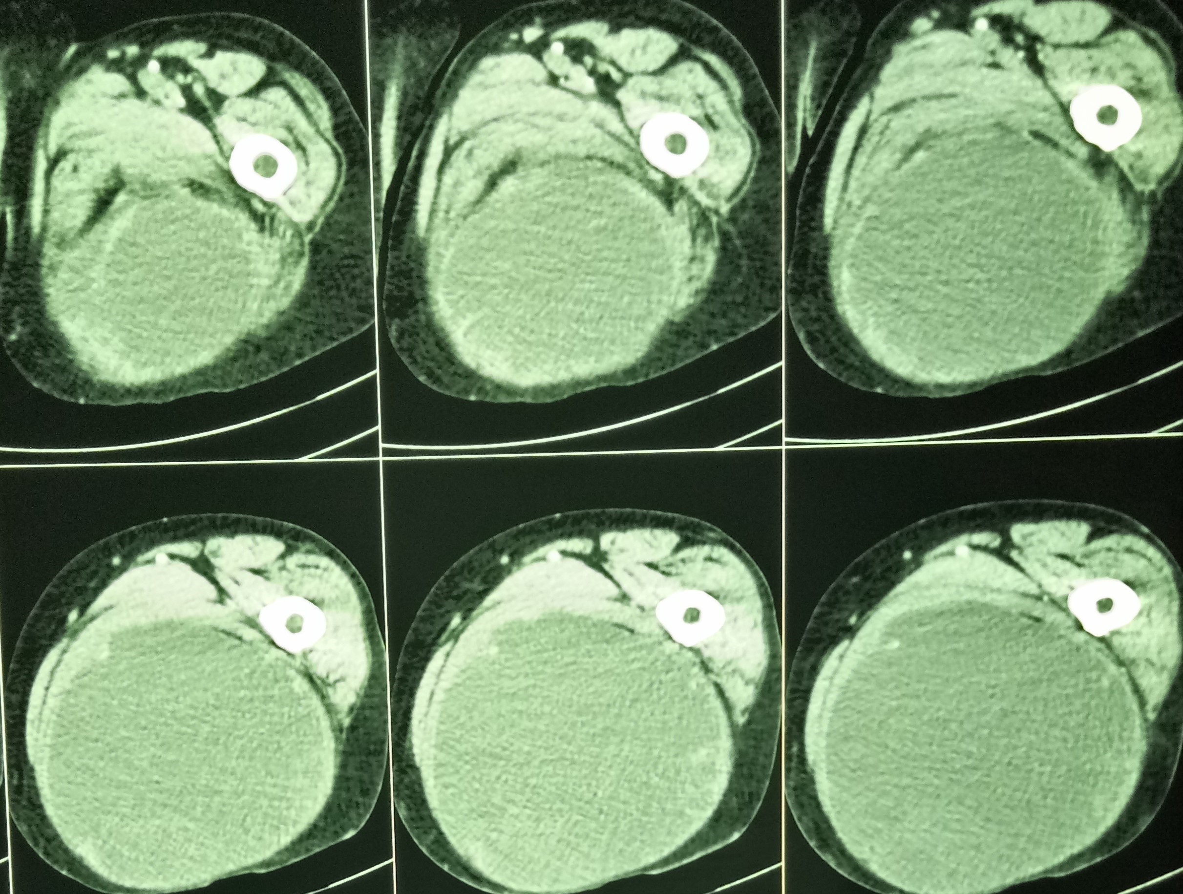
An atypical Fournier’s gangrene had resulted in perianal sepsis. The patient, a 50 years old male, first presented with pain and tenderness in the scrotum. There was a palpable crepitus in the scrotum – pneumoscrotum. CT scan showed a lot of gas in the scrotum.
The patient was treated with antibiotics and the pneumoscrotum gradually resolved but the patient developed a soft urethral stricture which was dilated.

Also he developed a perianal abscess which was drained. However, this eventually ended up in a complex horse-shoe fistula which was excised.
22.4.19
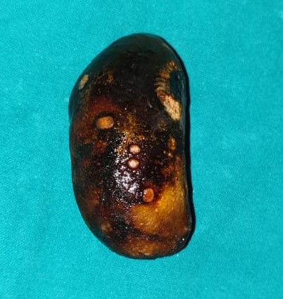
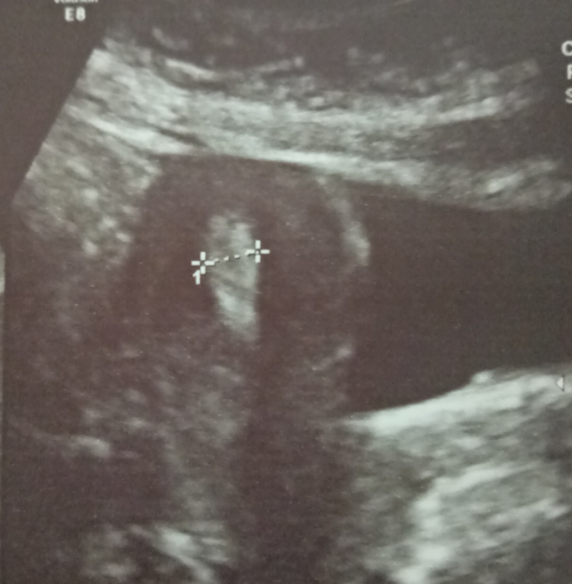
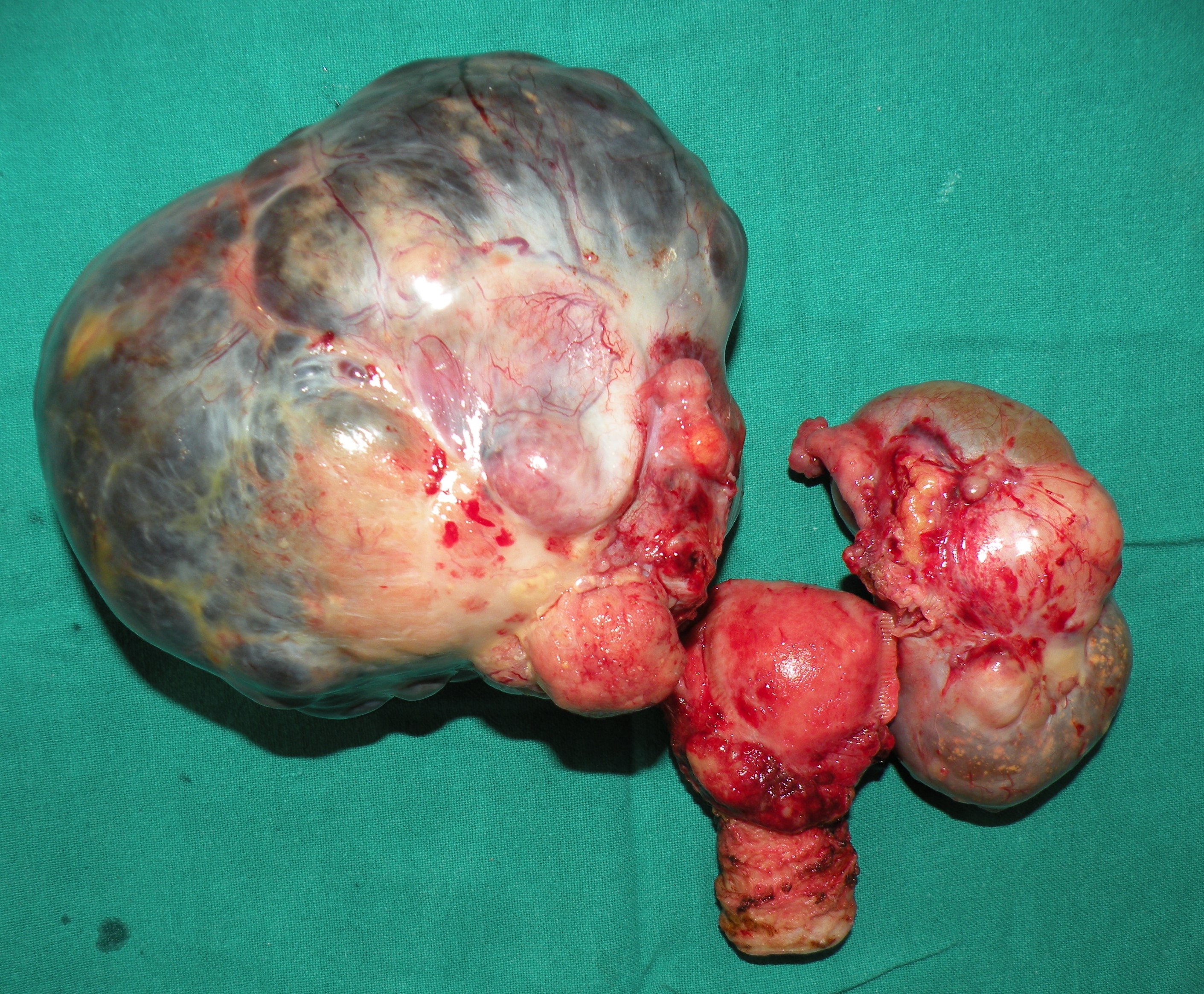
A supposed hydrocelectomy had to be converted into orchiectomy. Dr jagbir’s case. Hard testicular tumor with necrotic areas.
6.6.19
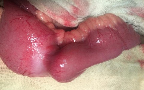
Called for strangulated right inguinal hernia in a 70 years old male Dr Vikram’s case). REEA of about 6 inches length of gangrenous ileum had to be done.
21.6.19
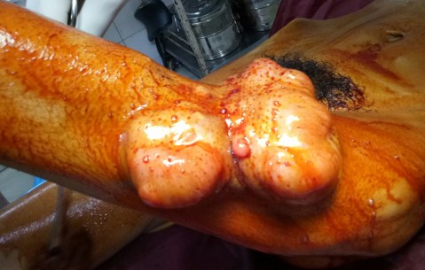
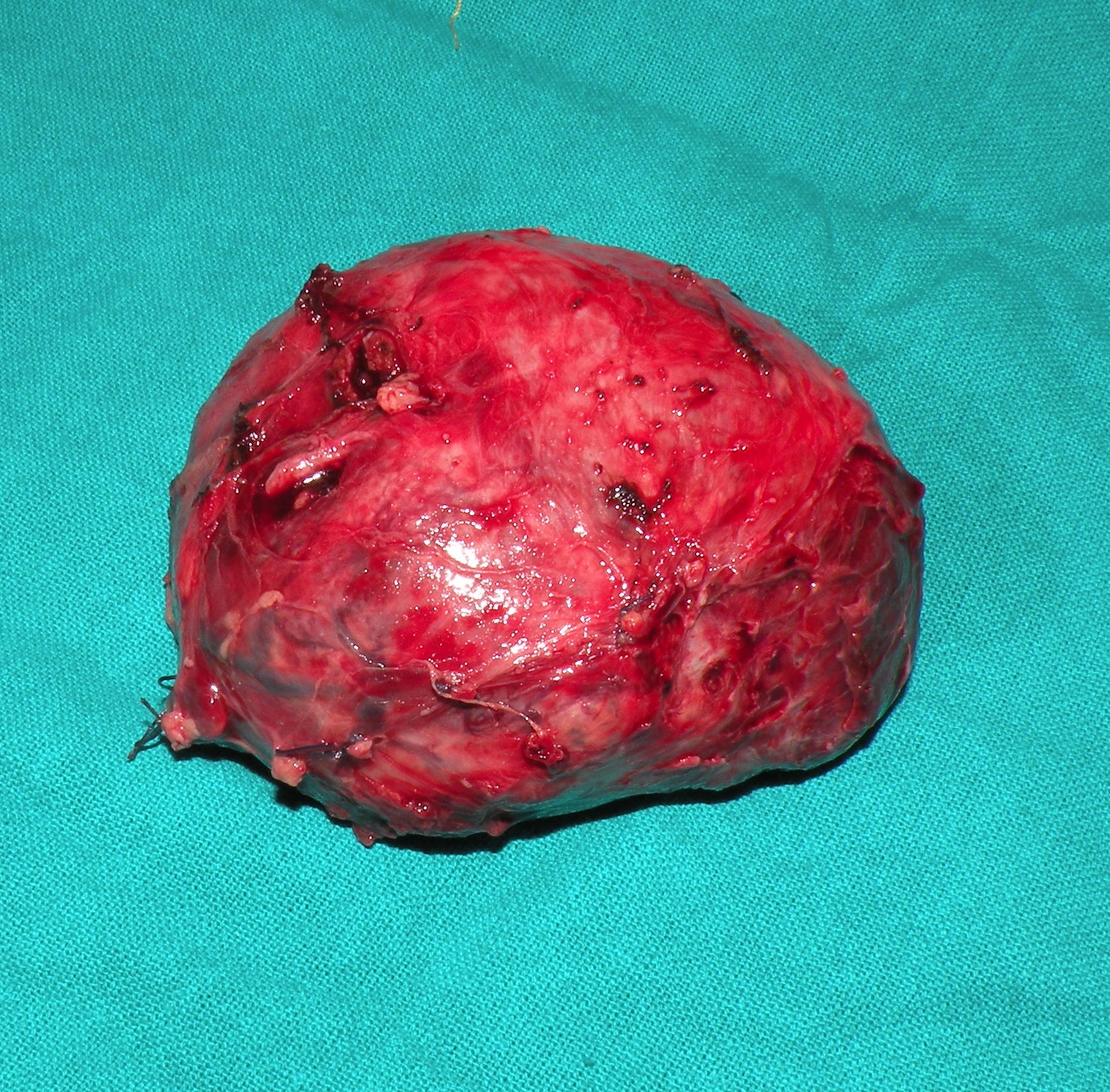
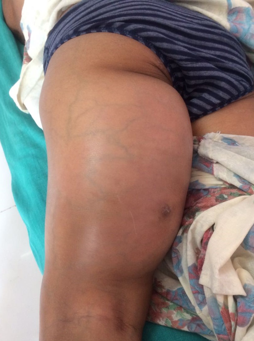
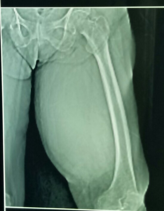
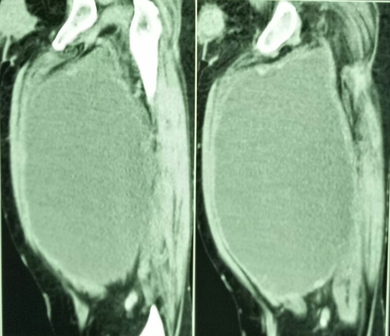
A huge recurred dermatofibrosarcoma of the left thigh in an elderly female was excised followed by skin grafting of the defect.
25.6.19
Sacrocolpopexy for vault prolapse. Previously had laparoscopic hysterectomy 20 years back at Doraha. (Dr Arora’s case).
26.6.19
Nephrectomy for non-functioning left kidney (due to stones of many decades standing) in a 68 years old female from himachal pradesh.

18.7.19
Sigmoid colon cancer resected with colorectal anastomosis. the adherent ileal loops as well as the adherent part of anterior abdominal wall excised too along with the colon cancer. A colorectal anastomosis and an ileo-ileal anastomosis had be made.
19.7.19
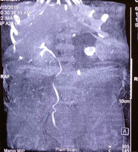
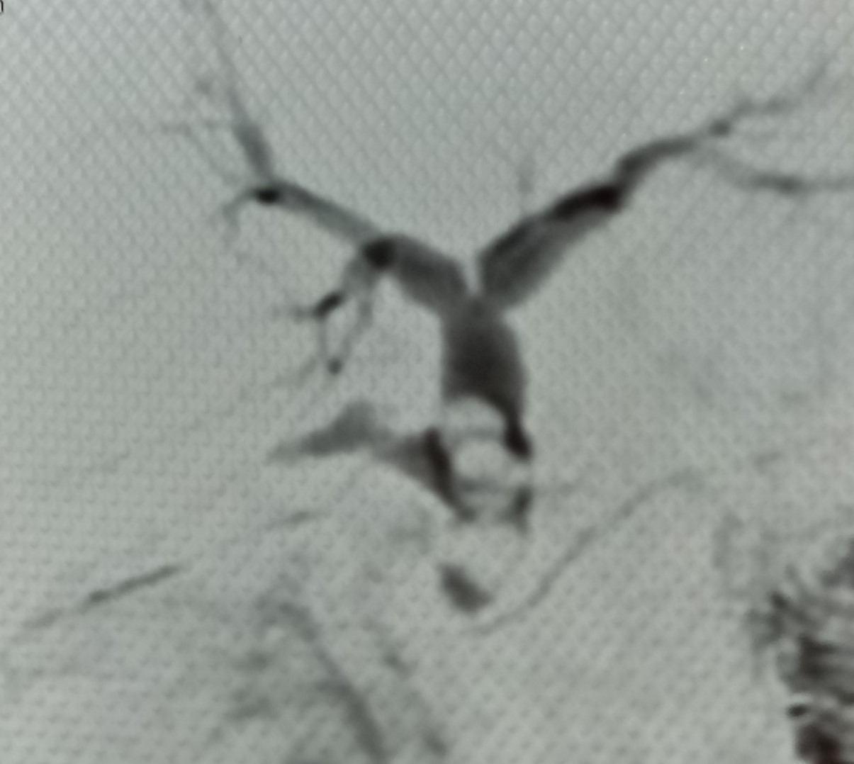
Laparotomy for recurrent episodes of small bowel obstruction in a 58 years old man. At operation, a stricturing tumour of the ileum was found. This was excised and EEA performed. Biopsy report was GIST.

8.4.19
A hefty tall 55 years old with opium addiction and depression (on treatment with anti-depressants) was hospitalized with acute abdomen and was found to have acute cholecystitis and a big distended thick walled gallbladder with a huge stone (around 5 cm) in it. He also had fecal impaction which was treated with enemas. Laparoscopic cholecystectomy was not considered and an open operation was planned. Even then, the big empyema and the difficult dissection of the Calot’s triangle took more than 2 hours.
Photos clicked by Dr Jasleen.

13.4.19
A similarly hefty and tall male patient, also an opium addict, underwent a similarly difficult procedure. However, the gall bladder and the stone were not so big and so the laparoscopic cholecystectomy was comparatively an easier option. Also, this patient was not depressed and psychotic like the previous patient and so was not difficult to manage postoperatively.
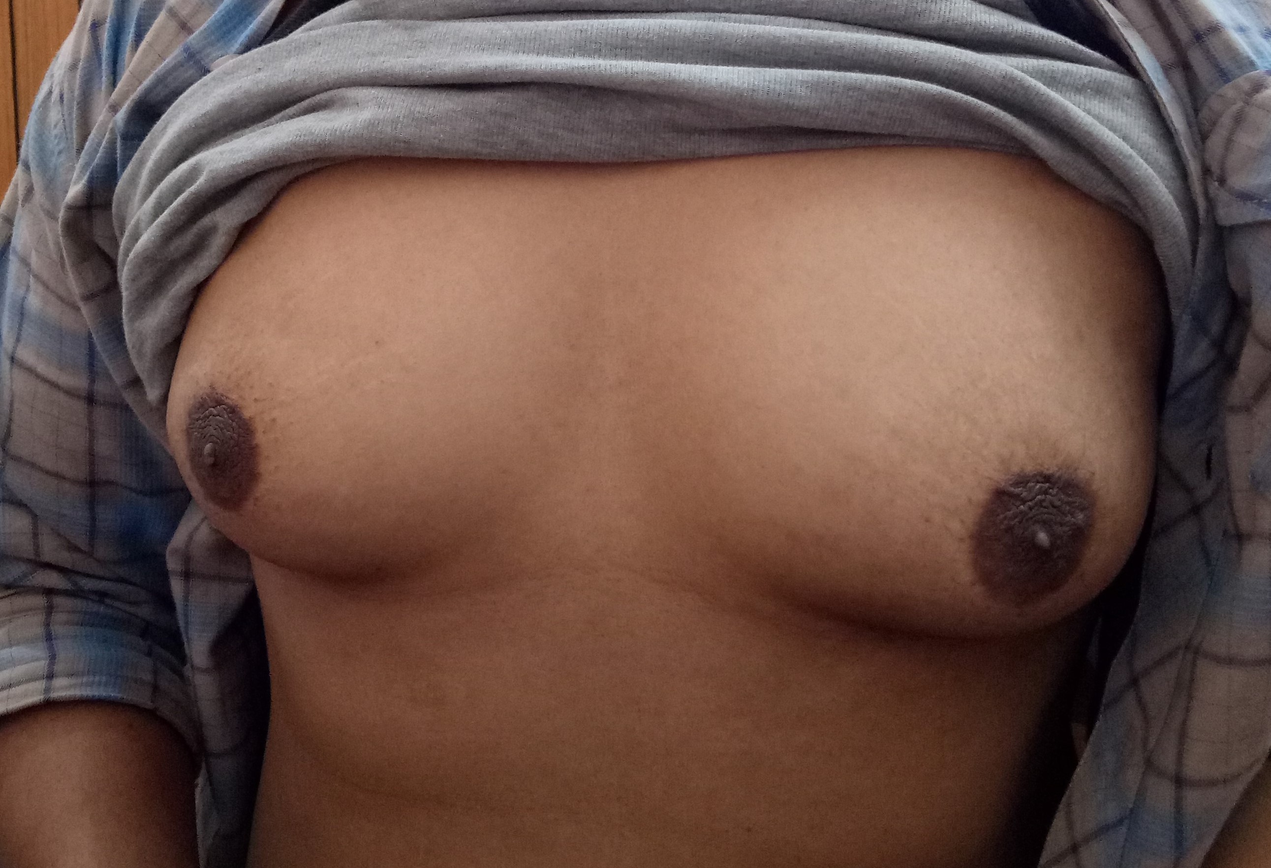
Gynecomastia
Posted on: August 25, 2019
6.4.19
A young fit man (had been obese and now into fitness programme) with big bilateral gynecomastia. Excised like bilateral mastectomy under GA.
27.3.19
A Nissen fundoplication was done in a 55 years old obese female in an open manner because of the lack of liver retractor.
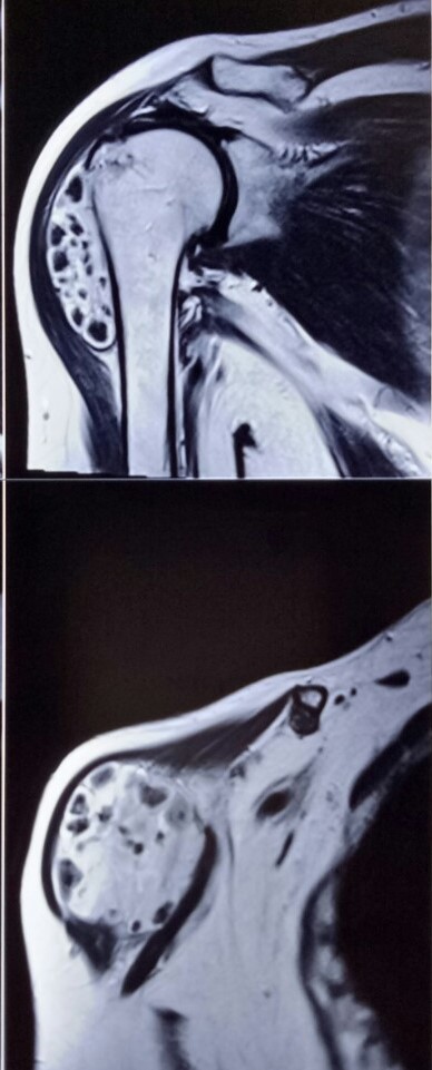
1.4.19
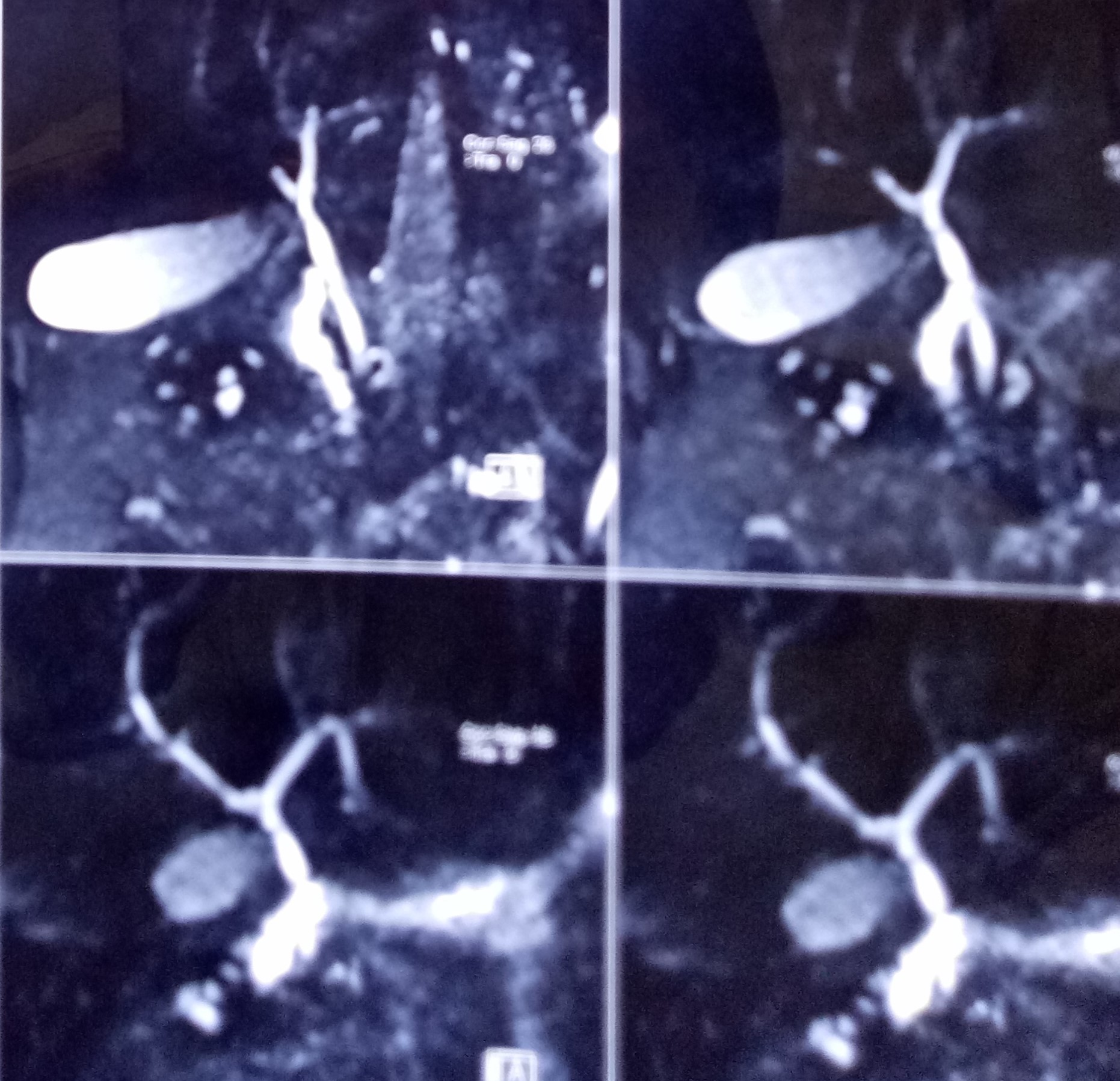
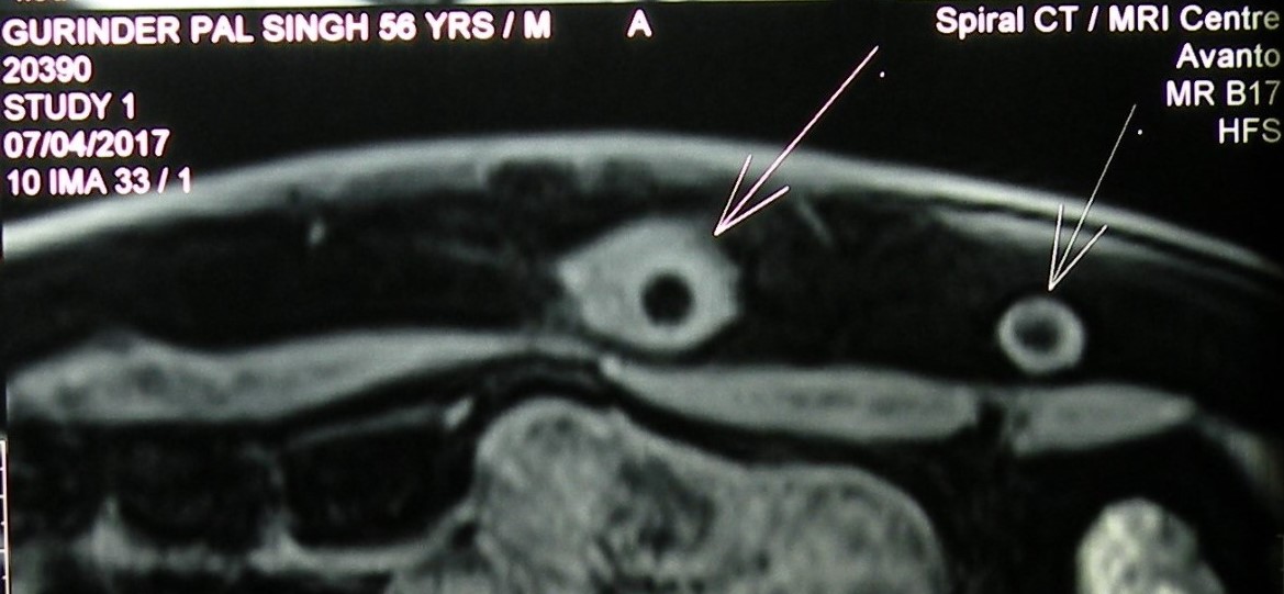
A 62 years old lady had a long-standing swelling over the right shoulder. MRI showed some adhesions with the upper end of the humerus. Was excised in toto. Biopsy report was hamartoma.


6.3.19
A young man (26 years old) had an appendix mass about 2 months back and was posted for lap appy. At operation there were still some adhesions with the anterior abdominal wall. One such band of adhesions, and an appendix epiploicae together were misinterpreted to be the appendix and so were removed. Later when the caecum was turned a little medially, the real appendix showed up from below and was removed too.!
6.1.19
An obese (>100 kg) lady, related to Dr Mrs Shalley, with a previous lap ventral hernia repair with a prolene mesh, needed a careful umbilical port entry under vision through a small scope brought in through Palmer’s point.
15.1.19
A young (35 years old) female, thought to have an easy lap chole, turned out to be a disaster, with the very shrunken gall bladder with a small stone in the fundus (which was possibly the whole gall bladder on retrospective analysis) having such dense fibrotic adhesions with the underlying bile duct so as to make them to look like a single structure. So the dissection was continued down for a couple of centimeters and the supposed neck of gallbladder ligated there. Bile continued to pour through the drain and the patient had to undergo a BDI repair in PGI.
Lesson learnt – in non-distended gall bladders on ultrasound, always have an MRCP roadmap before attempting surgery.
PET scan for breast cancer
Posted on: August 25, 2019
5.1.19
MRM for breast cancer, with PET scan showing axillary mets. 45 years old, c/o dr Jasleen.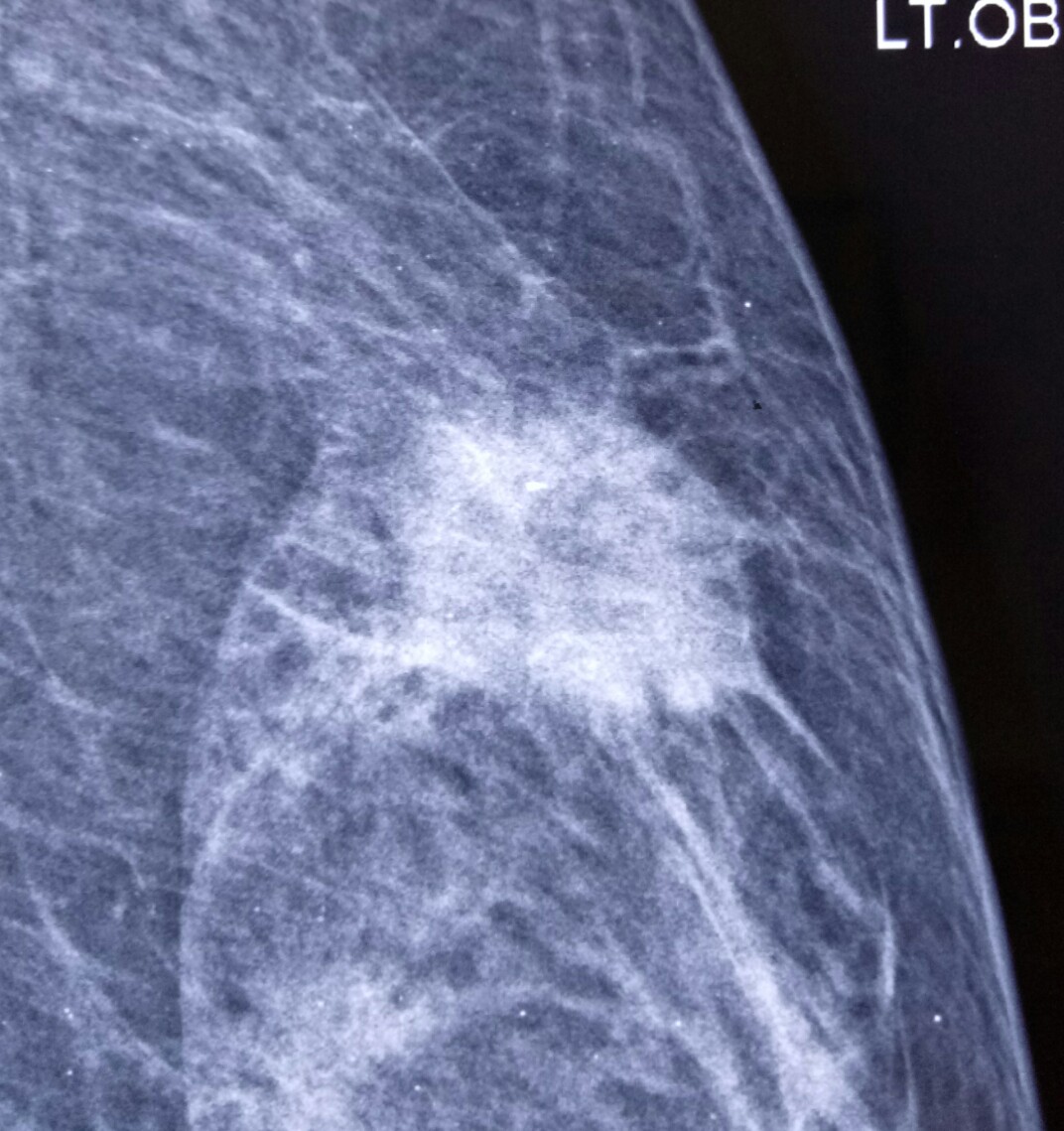

21.8.18
A 45 years old female had reportedly undergone an ERCP attempt to clear the CBD of stones but the stones could not be removed, and a biliary stent was placed. Another endoscopic attempt was made a month later, perhaps with an additional sphincterotomy (no records available, as usual) but ending up with biliary peritonitis (possibly due to a duodenal perforation) which was treated with US guided pigtail catheter drainage. The patient recovered and an MRCP on 2nd August revealed 4 big stones in the bile duct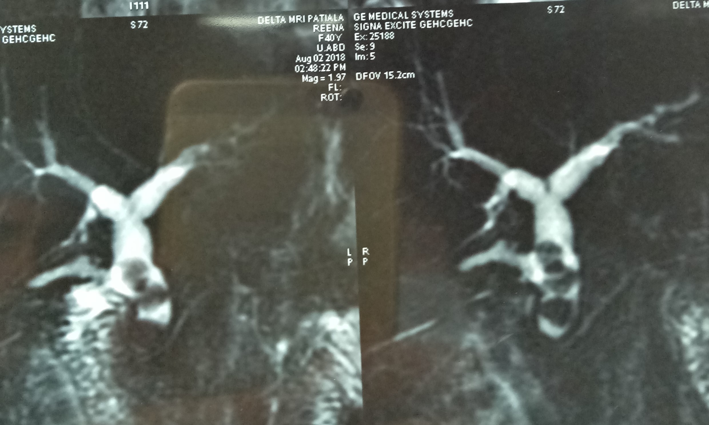
At open operation, the whole upper abdomen was found to be frozen in postperitonitis adhesions, and the CBD was located with great difficulty, and confirmed with needle aspiration. At choledochotomy, the CBD was found to be clear, with no stones. This was confirmed with choledochoscopy. A T-tube drain was placed. The gallbladder was not found. Postoperatively the patient did very well and remains well.
Why were there no stones in the CBD?? Possible they had passed out through the sphincterotomy!
20.10.18
An obese 40 years old lady, having consulted Columbia Hospital, reported for laparoscopic cholecystectomy. She had elevated serum alkaline phosphatase levels assessed repeatedly over a period of nearly two months, raising a suspicion of CBD stones. However, an MRCP showed no stones and a regular laparoscopic cholecystectomy was done.
Disulfiram implants, a big goitre, a bad splenectomy, a jejunal perforation by a trocar, an STS masquerading as an abscess, and a phyllodes tumor
Posted on: July 6, 2018
8.4.17
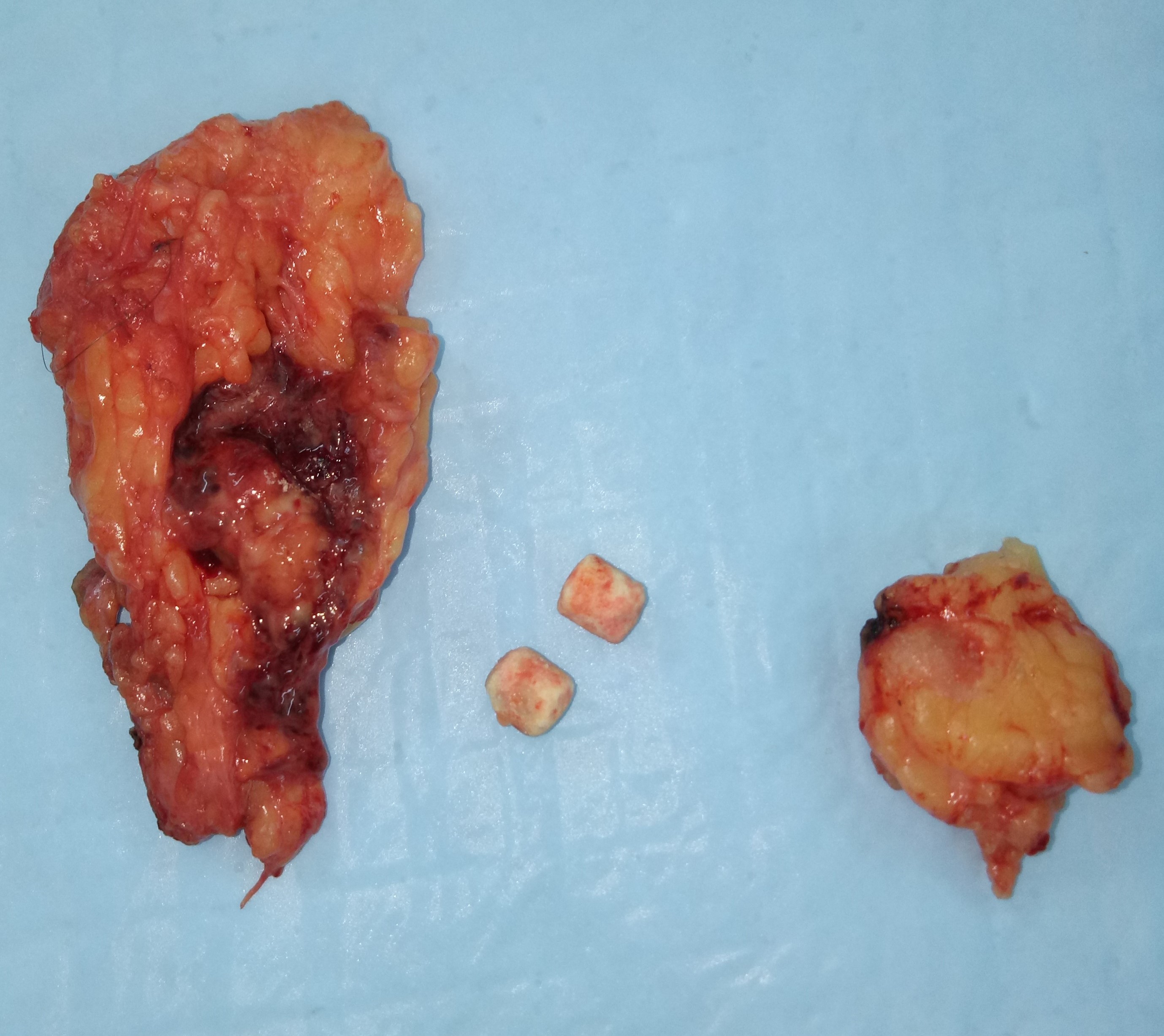
Excised two chronic abscess cavities that had formed around disulfiram tablets kept subcutaneously in the anterior abdominal wall in a 55 years old man, for his alcoholic addiction. Patient now reformed and normal and wanted removal of the implants which were causing pain.
I had never heard of such a thing as disulfiram implants. The present case had them implanted in Canada.
24.1.18
Cesarean hysterectomy for placenta accreta and percreta in an 40 years old lady with 12 weeks unwanted pregnancy. Very bloody surgery with large blood loss sending patient into hypotension (caused by the prolonged attempts by the attending gynecologist to manually separate the placenta), but fortunately ended well in the end. The D&C which was being discussed too in the management plan earlier by the gynecologist would have been disastrous.
25.1.18
A big vascular goiter in a 45 years old obese female. Right thyroid lobectomy took nearly three hours.
29.1.18
A lap chole was thrust upon me when Shashi Pathak w/o dr Pathak (microbiology) insisted on it even as she actually had no symptoms pertaining to gallstones. Actually turned out to be primarily suffering from depression for which was later treated by psychiatrists.
24.2.18
Splenectomy for shattered traumatised spleen 25 m. Bad and strange experience in the postoperative period when the patient continued to pour out litres of jejunal fluids through the nasogastric tube, and getting into huge water and electrolyte losses and dehydration. Explored on 1.3.18 to find acutely swollen and matted upper jejunal loops which were separated and washed clean. This did not help and another exploration was performed on 3.3.18 to rule out a mechanical factor in the distal loops. There was no such thing and the patient had to be kept on TPN for more than a week before he recovered. Inexplicable to me. Possibly, the poor anesthesia and poor relaxation caused a lot of bruises over the upper small bowel loops when they were covered with packs and retracted with lot of force. Also the abdominal packs given by the nurse were dry (not moist as they should have been). She said she never uses moist packs!
27.2.18
Another bad experience with a big incisional hernia (following pyelolithotomy) in an elderly 70 years old lady, repaired with a large mesh. Postoperatively developed a big seroma that took long time to heal.
5.3.18
Babaji from Dhanthal gurudwara presented with a hernia for which repair was done in Giani Lal Singh hospital.
6.3.18
A trocar perforation of the jejunum had to be repaired when called upon to do so by dr Vikram Tandon who was doing a lap chole.
7.3.18
Surinder bhahiji’s lap chole, went very well. Rinku had come all the way from New York to watch!
12.3.18
A presumed abscess of the thigh ( huge in size, diagnosed clinically and confirmed on CT scan) drained in an elderly ( 60 years old) female. The fluid was largely serous with lots of flakes. When examined after a month or so, the wound had partially healed but the cavity was filling up with a fleshy growth. Was referred to PGI where a high-grade soft-tissue sarcoma was diagnosed. She died soon after.
16 april to 17 may 2018 visit to America and Canada.
19.6.18
A lap chole for polyp and sludge in the gall bladder.
2.7.18
A big phyllodes tumor in the left breast of a young girl (18 years old) excised.
23.6.17
23 years old hefty young man had a hard small tumor of hard palate. Excised with cautery. Biopsy was benign pleomorphic tumor. Unfortunately developed a small palatal fistula possibly due to overuse of cautery. Had to be repaired by dr Harsimran, the ENT surgeon.
29.6.18
An epulis on left lower gingiva excised from a 55 years old lady turned out to be malignant. Referred to Tata cancer center Sangrur where it was re-excised and the patient remains well till date.
26.7.17
A gangrenous gall bladder excised laparoscopically with great difficulty due to frozen Calot’s triangle. Patient 56 years old male and related to sarpanch of village Chouhat.
10.8.17
Ovarian cancers bilateral excised at a TAH bSOP (dr Jagga’s case).
23.8.17
Attended the free medical camp held by Sarbat da Bhala trust in central jail to examine prisoners.
7.9.17
A badly neglected case of scrotal and testicular gangrene in a young man of 24 years ( referred by dr Sachdeva) explored and the testis had to be removed. Scrotal debridement and closure performed. Healed well.
23.9.17
An open CBD exploration (after failed ERCP attempts) and stone removal along with open cholecystectomy performed after a long time. This appears to me still to be a very satisfying procedure.
1.11.17
Joined VMHC (Vardhman Mahavir healthcare) as part time surgeon for 2 hours (9 to 11 am) everyday except Sunday.
5.12.17
A pilonidal sinus excised and closed in a young teenager male, only to get the wound infected after removal of sutures after 2 weeks and then for the wound to take more than 2 months to heal.
28.12.17
Fournier’s gangrene badly infected and the patient (65 years old man) septic going into MOD, shifted from some delhi hospital, underwent thorough debridement and survived. Scrotal skin could be closed several days later.
