15.12.09
La chole: ward worker, obese hypertensive lady 55 yr old.
Lap chole: 65 F admitted last week with gallstone pancreatitis which had resolved by now. Readmitted 25.12.09 with severe abdominal pain.
18.12.09
Lap repair of paraumbilical hernia.
Lap varicocelectomy 20 M.
19.12.09
Irreducible direct left inguinal hernia which had sigmoid colon nearly strangulated. 60M. Cord divided and mesh repair done.
20.12.09
(winter vacation 20.12.09 to 2.1.10).
TAHbSOP with sacrocolpopexy and mesh fixation of rectum to sacrum too for rectal prolapse. 65F from the hills with uterine and rectal prolpase. Had postop SBO due to adhesions for which laparotomy and adhesiolysis had to be done.
22.12.09
TLH for big fibroids made handling of uterus difficult. Took nearly 3 hours.
23.12.09
lap appy 25M, easy.
TEP for direct left inguinal hernia. 25M. Neat despite pneumoperitoneum and despited avulsion of inferior vascular pedicle which was simply coagulated and divided.
24.12.09
Laparotomy for suspected appendicitis which turned out to be biliary peritonitis in a 6 years old male child. The cause of bile leak traced to CBD the anterior wall of which had necrosed (cause??)and oozing bile. Repaired with cystic duct flap over a T rube.
28.12.09
TLH for huge uterus due to huge fibroids reaching upto above the umbilicus. Took nearly 5 hours. The uterus had to be cut into 4 pieces to be removed per vagina (morcellator not available).
28 and 29 november
Went to Kasauli with Dr Hardip Singh Mann and joined other classfellows of the 75GOMCO batch for a get-together. Stayed overnight and came back next day. Met old friends – dr Dharam Pal, Dr Tarsem Rattu, Dr Ravi Dawra, Dr Brij Kishore, Dr Bhucho (Keshav Garg), Dr Zora Singh, Dr Sohan Singh, Dr Gursharan Malhi, Dr Surinder Gupta, Dr Harjit Singh (braather), and many more.
30.11.09
Wide local excision (a near mastectomy) of an indeterminate mass despite 2 FNACs and mammography in an 85 year old female (an ex health worker – ANM). HPE- IDC. Started on tamoxifen.
A potential disaster averted just in time – lap chole in a 65 year old male, very difficult procedure. Liver firmly stuck to abdominal wall and absolutely immobile. Thick walled gall bladder with a very difficult grasp. A very easily dissected CHD, misidentified as CD, was clipped and about to be divided, when better sense prevailed, clips removed and a further dissection revealed a very short and thick cystic duct which was ligated with extracoporeally tied vicryl. Stormy postoperative period with abdominal distension and vomiting, the NG tube poured greenish output for a couple of days and then everything settled!! Could never tell what happened!!. A postoperative US scan and LFTs were normal.
1.12.09
Units of surgery department reshuffled after retirement of Dr Karam Singh. Mine will be 4the now, ward 3.
4.12.09
Microdochectomy for a single duct discharge from left nipple. 30 F, a principal of a school. Biopsy showed a well-formed papilloma.
9.12.09
Seminar on early detection of breast cancer.
11.12.09
A recurred swelling left forearm near the lower end of ulna. Presumably lipoma, turned out to be a swelling arising out of the tendon sheath of the extensor of the little finger, the ulnar artery pulsating close by. Excised under GA, preserving the tendon.
20.11.09
Laparoscopic excision of a big paratubal cyst (6 cm). Rather than the cyst alone, the whole mass of the cyst, tube and ovary was excised at the request of the attending gynaecologist. The other tube also coagulated with bipolar diathermy and cut (patient’s demand).
23.11.09
Carcinoid ileum – Right hemicolectomy for a presumed cancer caecum. A big mass palpable in RLQ of abdomen in a 20 years old female!. Getting obstructed with solid feeds, so kept on liquids for some days. CT showed mass in the caecum and ascending colon. Cut section showed mass in wall of terminal ileum rather than the lumen of caecum. Turned out to be carcinoid on histopathology and immunohistochemistry.

5.11.09
1. Big hydatid cyst right lobe of liver pressing on gall bladder and bile ducts, leading to elevated bilirubin levels. Laminated membrane excised in toto in one piece, and cavity partly obliterated (capitonage) and then filled with omentum. No drain.

2. Laparotomy for a mass lower abdomen and a vesicoenteric fistula, presumed to be a desmoid of abdominal wall. 17 M emaciated with history of blunt abdominal trauma, followed by history of urine contamination with faeces. At laparotomy, dense mass in lower abdomen with underlying loops of bowel adherent with it. Opened accidentally (and repaired) the transverse colon loop here while opening the abdomen. Painstaking dissection identified the proximal small bowel loops but the distal ileum, converted into a mass which was adherent to the bladder, had to be excised and EEA done. The bladder fistula was a small one and was closed.
8.11.09
30M taken up for appendicectomy thru small RLQ incision had to be converted into upper midline laparotomy on finding turbulent fluid in the peritoneal cavity. A DU perforation closed.
12.11.09
Two difficult lap choles:
1. Tripat Chand 72 years old male from Barnala (referred by Dr Surinder Garg and also known to Dr Avinash Gupta, AP orthopedics). Had a mass covered with omentum as if it was covering the gall bladder but on some dissection, a non-adherent gall bladder could be visualised behind this mass. Routine cholecystectomy was done with some difficulty due to the mass coming in the way. The mass (possibly with transverse colon or distal stomach) to be investigated later with CT and other investigations.
2. Post pancreatitis (gallstone) lap chole. 35F. Thick walled cystic duct, endlooped and ligated.
16.11.09
Laparoscopic ureterostomy (Dr Sukhpreet’s case) for a presumed stricture or radiolucent stone right midureter in a 30 years old female with pain and right hydronephrosis and hydroureter ureter above the stricture. Transperitoneal laparoscopic approach easily identified the bulge in the right ureter just below the caecum and over the pelvic brim. Peritoneum over it opened and ureter opened longitudinally over it with hook cautery to reveal the curled up stent in it but no stone. The thick wall of the stricture was further opened up and the stent pushed up into the renal pelvis. Uterer closed over it with 3-0 vicryl and the peritoneum too was closed over it.
17.11.09
A most difficult lap chole. 65 F obese patient. Tense empyema aspirated to begin with. The neck of gall bladder found to be very firm as if it contained a hard impacted stone in it. This neck was opened up to find a very thick walled abscess here. Dissecting below this could have been dangerous, so fundus first dissection was done upto this point and a subtotal chole was done. Three big stones removed from within the opened up gall bladder. These were broken into pieces and removed piecemeal. Then the gall bladder walls were also cut into several pieces and removed . Drained.
18.11.09
A difficult TAHBSOP. 40 F with previous unsuccessful attempt at Sirhind. The problem was the big fibroid with posterior wall of uterus. This was stuck in the pouch of Douglas, and pressing upon the displacing the right ureter.
29th october to 31st october
Attended the annual conference of International college of surgeons (indian section) held at GND university by Dr US Dhaliwal and team.
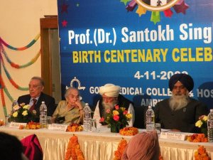
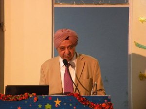
then again to amritsar on 4.11.09 to attend the dr SS anand’s birth centenary celebrations at Govt Medical College campus.

25.9.09
3 difficult cases –
1. TAHSOP
2. Appy lap : 30 M handicapped (RLLPPP) with h/o burst mass appx 6 months back. Tip of appendix still badly stuck retroperitoneally.
3. Laparotomy for neglected intestinal obstruction. Poor young (30) female, with previous 2 LSCS. At op, gangrenous loop of jejunum (thick omental band in pelvis from previous CSs) resected and EEA done.
27.9.09
Lap chole in 55 M (brother of Dr Ravinder Singh ex-prof orthopedics), difficult. Thick walled gall bladder and the thick short CD end-looped.
5.10.09
Lap chole converted to open. 70F with previous ERCP and ES for CBE stones. Dense adhesions with stomach. Calot’s triangle not clear even at open. Big stone impacted in neck removed and the neck ligated.
7.10.09
Laparoscopy for right ectopic tubal pregnancy which had already aborted thru fimbria. Thus tube could be saved after securing haemostasis from the fimbrial margins.
17.10.09
diwali
19.10.09
seminar on small bowel tumours.
20.10.09
Tried removal of a big chocolate cyst left ovary (dr jagbir’s patient) but had to convert due to the big size of the cyst (reaching upto above the umbilicus) and adhesions due to previous surgeries (CS and TAH). Even at open, was a tedious job.
22.10.09
Lap chole, day case, 52 F with acute cholecystitis, and thick walled gall bladder.
Nephrectomy (L): 30F with non-functioning kidney from long-standing PUJO. Calcified hydronephrosis. Retrospectively, could well have been done laparosocopically.


- In: operations
- 2 Comments
15.9.09
Meckel’s diverticulectomy 45M being operated for suspected appendicitis, actually had Meckel’s diverticulitis.

17.9.09
Burch colposuspension after TAH by dr Hans, colposuspension by me. Elderly obese lady. Used a mesh strip.
18.9.09
Cystogastrostomy and cholecystectomy. 45M with the cyst persisting for 6 months now.
7.8.09
joined GMC Amritsar. surgery unit 3 (with dr gulati)
9.8.09
2 lap choles in patiala:
day case lap chole 10 at MS surgicare and
one more.
11.8.09
Assisted Dr Sukha Singh at TAH at his clinic (Amrit hospital) in Amritsar. Elderly lady with atrophic uterus. ? indicated or not?. Postop had severe abdominal pain and distension and vomiting, frightening the surgical team but eventually settled in 3 days.
13.8.09
MBBS final theory class in old LT in Amritsar medical college. CHPS.
17.8.09 monday
MBBS final theory class, continued with stomach – PU. Reached at 7.30 am from Patiala in time for the class.
20.8.09
last of these classes in amritsar, as JDA went on strike after this.
21.8.09
JDA on strike, junior doctors in Amritsar lathicharged by police in front of the OPD.
29.8.09
day case lap chole 11. previous ERCP and ES and stone extraction. Dr Kaler’s patient.
30.8.09 sunday
Went to chandigarh with mr Bhalla and saw the minister Tikshan Sood for transfer. He consented.
31.8.09
TLH , satisfactory.
8.9.09
To Amritsar and back, after having been relieved from there after transfe back to Patiala.
09.09.09
Joined at Patiala GMC again. good date to remember.
Also to remember was the first laparoscopic nephrectomy for non-functioning hydronephrosis (R). Very satisfactory. Specimen removed in a bag. Enjoyed.
11.9.09
A big ovarian cyst laparoscopically excised and removed in a bag.
13.9.08
two difficult cases:
a huge man 25 years old but very obese , more than 150 kg. Varicose veins ligated at saphenofemoral junction (Trendelenburg) plus multiple phlebecotomies.
Anderson Hynes pyeloplasty in a 7 years old male child. Stent not available. Drained.
22.6.09
Day case lap chole number 6: for the first time, port site (epigastric) bleeding caused a nuisance, controlled afterwards (after removing the gallbladder) by two sutures with port-closure needle.
23.6.09
Lap chole day case 7: Asha verma 55F, w/o popular school teacher here. short and wide CD, endlooped.
7.7.09
A disaster: trochar vascular injury during first trochar insertion for the camera in a case of diagnostic laparoscopy at Sehat, injuring the aortofemoral junction on the right side, leading to shock and a big haematoma, called in to repair, but unsuccessful. Patient succumbed. Sad day.
9.7.09
Emergency call for caesarean hysterectomy. Patient in shock. Saved after a quick hysterectomy at Babbar’s.
18.7.09
Repair of extensive vaginal lacerations at vaginal delivery conducted by a dai.
20.7.09
Accessory duct at day case 8 lap chole; doubly clipped and underrun too. OK in the end.
21.7.09
Bilateral TEP for recurrent bilateral inguinal herniae. 65M, related to the mess contractor of doctors’ hostel mess.
22.7.09
day case 9 lap chole, 40 F thin lady. Easy and nice postoperative period.
27.7.09
Failed retroperitoneal ureterolithotomy.
3.8.09
Relieved from GMC patiala for amritsar.
Joining govt service again, and some surgeries (including an adult intussuception and a hymenectomy).
Posted on: June 21, 2009
24.5.09
Acute gangrenous appendix removed laparoscopically urgently at 10 pm (c/o dr parminder from barnala). Young 12 male child.
27.5.09
A twisted ovarian cyst clinically and radiologically diagnosed, excised laparoscopically. 15 years old female with severe unremitting pain for 5 days (c/o milkman). However ovary was seen separate from the mass, and therefore it was surmised the mass was arising from the tube.
1.6.09
12 male child with a preoperative diagnosis of varicocele, causing pain. At inguinal exploration, had an indirect hernial sac which was the most likely cause of pain. Herniotomy and ligation of varicocele done.
4.6.09
Joined GMC patiala again as professor after the notice of premature retirement given in february was allowed to be withdrawn by the government.
5.6.09
Laparotomy for intestinal obstruction of some days standing. Remained admitted with medicine department with lots of investigations – including CT and BMFT but not operated. At operation, had intussusception – ileoileal. REEA done. A tumour within the lumen seen on cut section of the specimen.
7.6.09
day case lap chole 5. postop fever upto 101, settled in evening.
11.6.09
TLH satisfactory and complete.
12.6.09
TEP repair of left inguinal hernia. Very satisfactory and clean at last. 50 M with small direct hernia.
14.6.09
TLH satisfactory. Obese female 45 y o with posterior fundal fibroid.18.6.09
18.6.09
Hymenectomy for imperforate hymen. 17 F with big haematocolpos and haematometra.
21.6.09
a possible CBD injury:
an easy looking lap chole became difficult. 35F. while creating the posterior window and dividing the peritoneum posteriorly, bile leak noticed, presumable due to accessory duct. The patient was later found to have a RHD injury. Sad.
