Archive for the ‘Uncategorized’ Category
Situs ambiguous(heterotaxia) with gall stones, an ileal carcinoid? and bad encounters with chronic liver disease
Posted on: October 11, 2013
9.9.2013
A lap chole converted due to impossible access (previous laparotomies for ileal perforation peritonitis, then closure of ileostomy + dense perihepatic adhesions).
18-20 September, 2013
Attended a SELSI conference at Srinagar. Good workshop showing hernias, lap ureterolithotomy, lap pyeloplasty, lap nephrectomy, lap CBD exploration.
23.9.13
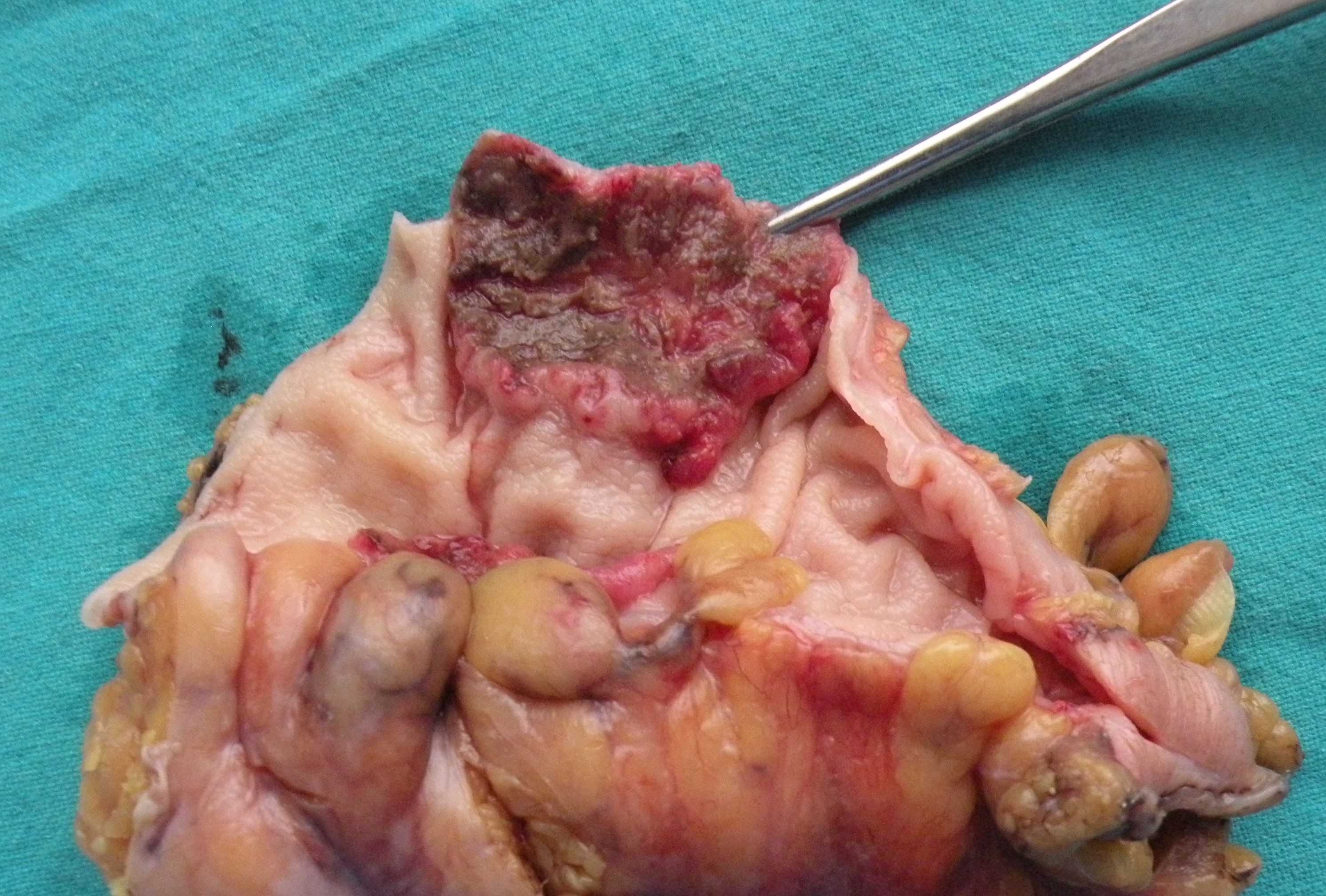
Laparotomy for long-standing history of intermittent colicky abdominal pain in a 40 years old male. diagnosed presumptively with tuberculosis of intestines, and treated with ATT without relief. A recent CT showed 2 ileal strictures. At laparotomy, the segment of mid-ileum bearing the 2 strictures was excised and EEA done. Biopsy surprise was carcinoid.
26.9.13
Encounter with CLD 1 – A very difficult lap chole ended up in conversion. CLD with previously low PTI and previously postponed several times. Presently all LFTs and PTI reported to be normal. A very thick-walled and adherent gallbladder dissection resulted in duodenal tear and prolonged oozing from liver bed. After conversion to open, bleeding controlled with pressure and suture of liver bed, and the duodenal tear repaired. But the patient ended up in ICU due to incomplete recovery from anaesthesia, and there, her LFTs deteriorated and she died on 5th postop day.
7.10.10
Encounter with CLD 2 – A 50 years old female admitted with acute cholecystitis, now settled. Previously known history of portal hypertension and bleeding varices controlled with sclerotherapy for last 2 years. LFTs in normal range now – Childs grade A. So taken up for lap chole and burnt fingers again. Big vessels all around the gallbladder and in Calots triangle. Opened up for bleeding in Calots area, not controlled by pressure laparoscopically. At open operation too, the bleeding from liver bed severe, suturing of liver bed led to further bleeding, compounded by a retractor injury to the liver bed. 2 abdominal packs and pressure controlled the bleeding, and patient closed with packs in place. The packs were removed 5 days later when the patient stabilized, but the packs were found to be getting infected, thought the bleeding had stopped. The patient continued to drain through the abdominal drain for many more days.
10.10.13
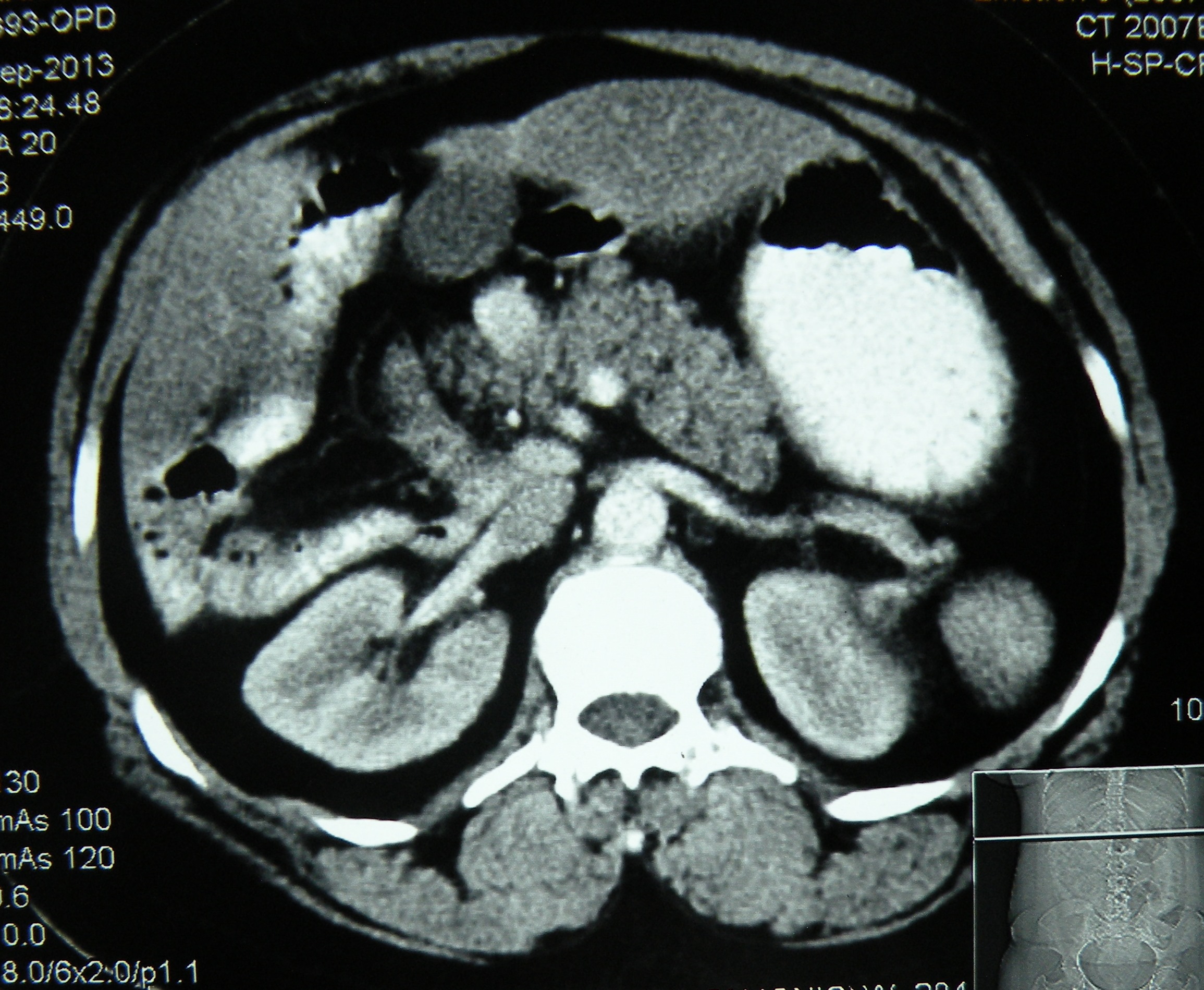
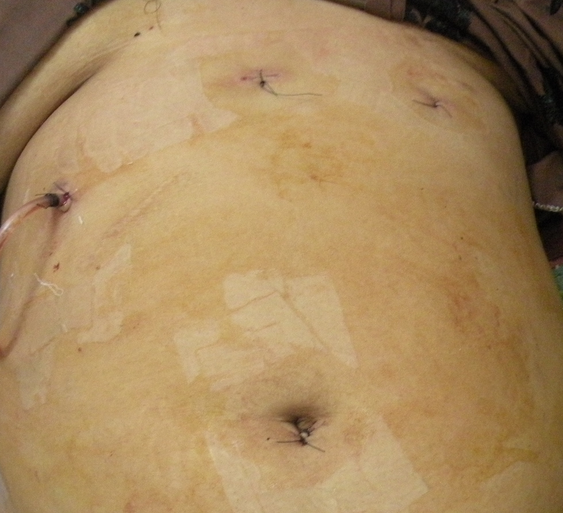
An interesting case of situs ambiguous (heterotaxic syndrome) presented with history of failed open attempt at civil hospital, Nabha to remove her gallbladder which could not be found by the surgeon. Imaging (US, CT, MRI) here showed a central liver, with gallbladder placed in the centre between the two lobes, polysplenia, truncated pancreas and dextrocardia. At surgery, the gall bladder was found just to the left of the falciform ligament. A difficult lap chole (due to dense adhesions with falciform ligament and omentum) was done, with the main operating port in the LUQ of the abdomen.

 published later in
![]()
| CASE REPORT Year : 2014 | Volume : 4 | Issue : 3 | Page : 180–182 Laparoscopic cholecystectomy in situs ambiguous Anoop Varma1, Abhinav Mahajan1, Mohinder Singh1, Gunjeet S Sandhu1, Navkiran Kaur2, |
A rectal cancer anterior resection, a breast cancer extirpation, a couple of difficult lap choles converted, and a variety of ectopic tubal pregnancies
Posted on: September 2, 2013
28.7.13
Ruptured left tubal pregnancy, lap salpingectomy with bipolar forceps.
29.7.13 A similar tubal pregnancy, similarly dealt with.
3.8.13
Right tubal ectopic this time, looked like a solid tumour inside of which were the products – could this be the result of methotrexate treatment which had been given to the patient?
5.8.13
Difficult lap chole (Dr Mohi’s patient 50 F), converted to open due to a big impacted stone in the neck of gallbladder and the calot’s triangle frozen.
8.8.13
Another conversion of lap chole to open, this time a 50 years old male patient admitted with acute cholecystitis last week. At operation, a thick walled empyema and Calot’s triangle could not be dissected. Had a stormy postop period. First 4 days normal , but had pain and distension of abdomen on 5th PO day. ERCP showed CBD stones, which could not be removed (?impacted), and CBD was stented. Then had pneumonitis which gradually settled with antibiotics.
20.8.13
Massive gangrene small bowel. A poor gardener, 50 years old, was admitted with 7 days old history of intestinal obstruction, kept on conservative treatment at Rajan NH, was found, at laparotomy, to have distension and gangrene of nearly whole of the small bowel, excepting nearly 2 inches of proximal jejunum and 2 inches of terminal ileum, which were anastomosed after resection. Was referred to PGI for TPN and further care, then was lost to follow-up.
A chronic ectopic ruptured tubal pregnancy, forming a solid mass, removed piecemeal.
2.9.13
MRM for an advanced (skin fixity) cancer of left breast. Lymph nodes (not palpable clinically) were a fixed mass adherent with axillary vein.
Anterior resection for a rectal cancer middle rectum (60 M, dr Jagga’s case). Shocking start of the operation because of the nick (with cautery) to the left iliac artery while beginning the mobilization of the sigmoid! Closed with 4-0 prolene. Then smooth sailing. Total mesenteric excision and stapled anastomosis of the descending colon to lower rectum.
12.6.13
Thick-walled empyema of gallbladder, removed piecemeal.
19.6.13
Big perforation of duodenal ulcer in a 30 year old smoker. Could not be closed primarily, only plugged with omentum.
21 t0 24 june, 2013
Lots of operations performed in a free medical camp at Baru Sahib, Himachal Pardesh: 7 open cholecystectomies, 5 inguinal hernia repairs, 3 paraumbilical hernias, and 2 hydroceles.
11.7.13
Complications arising out of a difficult lap chole (c/o staff nurse, OT). Thick walled empyema and fibrosed Calot’s triangle, ligated at infundibulum. Removed piecemeal after more than 2 hours of effort. Had bleeding from omentum postop that had to be ligated at laparotomy.
17.7.13
Priapism of 4 days duration in a 70 year old man!. Partial detumescence achieved by aspiration of corpora cavernosa and by injection of saline norepinephrine solution.

18.7.13
An attempted TEP (60 years old man) failed because of intraperitoneal entry in the beginning itself, converted.
19.7.13
Court evidence at Pathankot, went via Jalandhar.
25.7.13
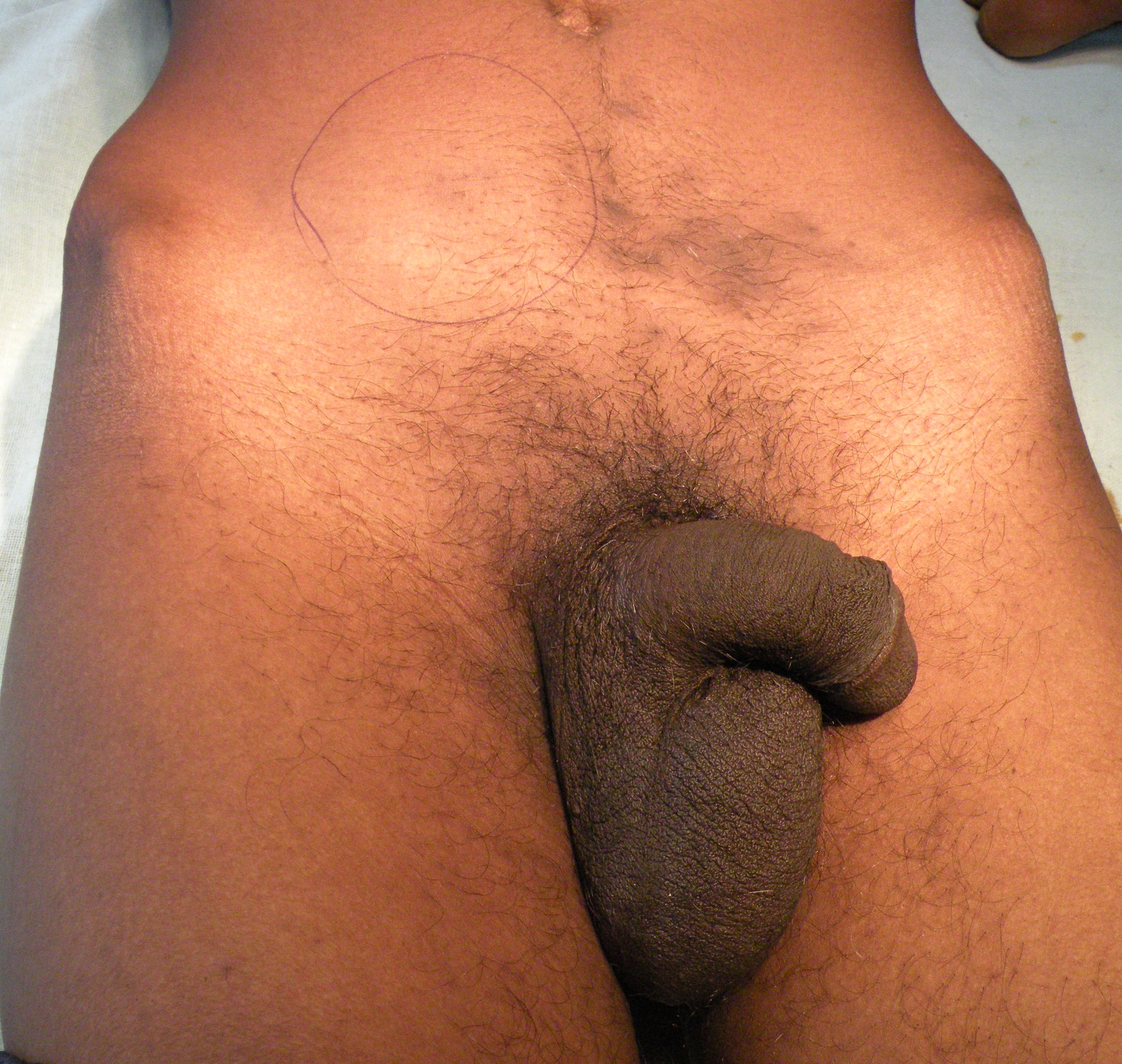
A tumour of undescended testis on the right side in a 23 years old man. Excised at laparotomy.
 <
<


Appendix stumpitis, branchial cyst, mesenteric fibromatosis, and a femoral hernia
Posted on: April 22, 2013
5.4.13
30 years old female, thought to have pain right lower abdomen because of a TO mass (on US), turned out to have no such mass, but a stump of appendix (left after previous open surgery) – removed laparoscopically.
11.4.13
A large branchial cyst in a 15 years old girl, excised. Contents purulent, hence the pain and sudden enlargement recently.
 <
<
 </a
</aLaparotomy for a solid mass in 12 years old boy – turned out to have a solid big mass from mesentery, attached to ileum, which had to be resected along with the mass. The biopsy report was mesenteric fibromatosis.
 >
>
Case Reports in Surgery
Volume 2013 (2013), Article ID 569578, 3 pages
http://dx.doi.org/10.1155/2013/569578
Mesenteric Fibromatosis Presenting as a Diagnostic Dilemma: A Rare Differential Diagnosis of Right Iliac Fossa Mass in an Eleven Year Old—A Rare Case Report
Abhinav Mahajan, Mohinder Singh, Anoop Varma, Gunjeet Singh Sandhu, Malwinder Singh, and Rupesh Nagori
22.4.13
Femoral hernia, strangulated, in a 70 years old female. Could not be reduced through lower incision. A lower midline laparotomy added, loop of strangulated terminal ileum released, and an end to end anastomosis made after resection. Hernia repaired from below with ethilon 1-0 interruptted sutures.
GB agenesis, vasovasostomy etc
Posted on: February 18, 2013
15.1.13
A lap chole in a 60 years old man with empyema tested skills. GB cut open and removed in pieces after removing the stones. The gallbladder ligated at its neck without identifying the cystic duct which could not be identified. The neck ligated again with preformed catgut loop (ethicon), but leaked bile for some days through the drain, eventually drying up.
28.1.13
MRM of mother-in-law’s cancer (medullary) of breast. L nodes reported negative.
4.2.13
Abdominoperineal resection for an anal canal cancer which did not respond to Nigro’s chemoradiation therapy. 60 years old man. The perineal wound had apparently healed nicely when he was discharged after 25 days. However, a week later, the perineal wound gaped (previous radiotheray, delayed healing?), and was resutured.
5.2.13
GB agenesis in a 25 years old male (bhukki addict) presenting with pain upper abdomen and US reporting gallbladder stones. No gallbladder could be found, after removing adhesions of omentum and colon mesentery in the gallbladder fossa.
11.2.13
Vasovasostomy in a 50 y o man who wanted to have children again.
18.2.13
Open CBD exploration in a 50-year-old female (relative of Dr Rama) for a big calculus (primary) in the CBD. She had an earlier endoscopic sphincterotomy but the stone could not be removed, and a stent had been placed. Today, the single big stone was removed from CBD. No stone was found in the gallbladder removed.

13.12.12
TEP repair of inguinal hernia on a 60 years old man, the inferior epigastric pedicle got detached but no further problems with dissection.
18.12.12
Held a clinical meeting in ME cell of the college, wherein two rare presentations of tuberculosis of the vertebral spine were discussed. The cases were presented by Dr BL Bhardwaj, professor of medicine.
15.1.13
A very difficult empyema of gallbladder in a 60 years old man. Lap chole by opening the gallbladder first, emptying it of stones, and then fundus first dissection, and then ligating at the neck.
19.1.13
Herniotomy in a one year old female child with right inguinal hernia. The contents were the ovary and fallopian tube, saw this for the first time.
21.1.13
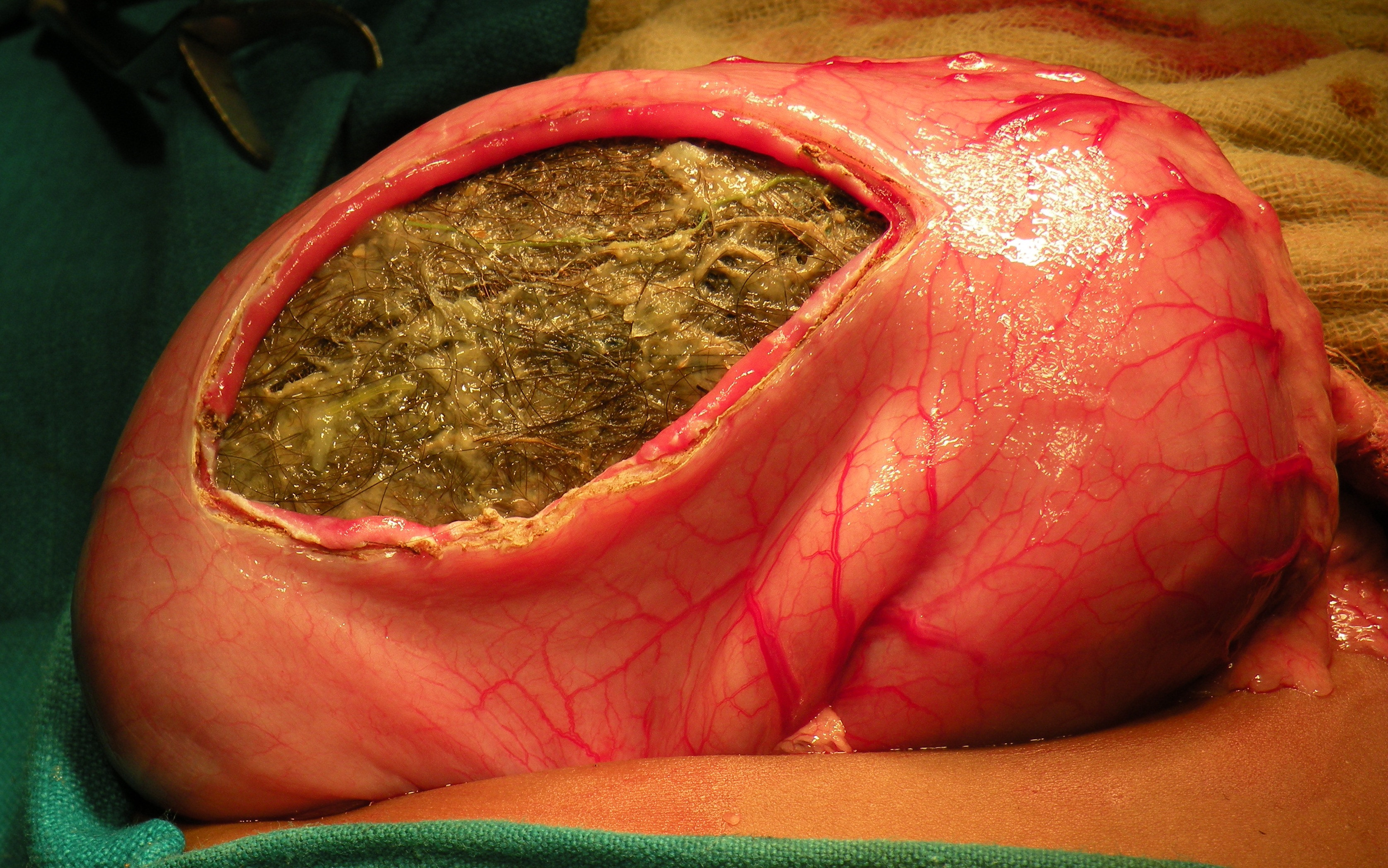
Trichobezoar removed from the stomach of a 5 years old female child.

Plexiform neurofibromatosis
Posted on: November 5, 2012
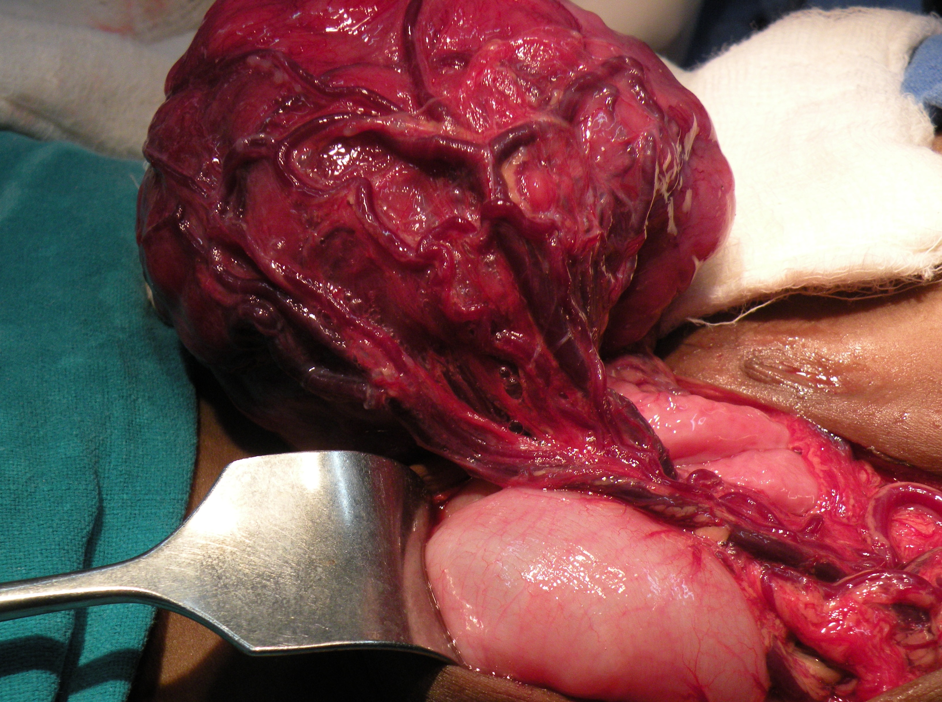
A huge neurofibroma, a part of von Recklinghausen’s NF, excised from the back of a 25 years old lady.

25.10.12
Re-exploration laparotomy for an obstructed gastric outlet, after laparotomy 3 weeks earlier by residents, in a young (20) male, with transection of duodenojejunal junction following trauma in a RTA. After the duodenal continuity was restored with a sutured anastomosis, a GJ had been added!. The patient continued to have more than 500 ml of bilious NG aspirate, and on removing the tube once, had some bilious leak from the abdominal wound, suggesting anastomotic leak, so the tube was put back in. At operation, the the DJ anastomosis was found to be strictured, and was dilated through an enterotomy with foley balloon and metal dilators, and the GJ was converted to RYGJ.
1.11.12
At lap chole, measured the dimensions of a Rouviere’s sulcus with the help of a feeding tube marked in centimeters, as a part of studying the anatomy of Rouviere’s sulcus as seen during laparoscopic cholecystectomy.

NCASICON at jalandhar – attended for two days : 8 and 9 september. Saw dr Chanjiv and others there.
13.9.12
Open prostatectomy for a huge (150 g) prostate that presented with hematuria. Postoperatively, developed clot retention, and had to be reopened to clear the clots.
Undescended testis in a 2 years old child- no testis found – anorchia.
20.9.12
Closure of ileostomy for a patient of Crohn’s disease who had presented with obstruction and residents excised a segment of ileum and had done an ileostomy. Patient on azathioprine by his GP.
Revision of GJ into Roux-en-Y GJ. An older patient who had presented in 2006 with GOO and had TVGJ performed. Now continued to have pain and vomiting intermittently. At operation, had stenosis of both limbs of GJ, which was revised into Roux-en-Y GJ. Did well thereafter.
21, 22 sept
Attended SELSI hernia conference at Gwalior along with Dr Sushil Mittal and his team.
28.9.12
Abdominoplasty (assisted dr GP singh) for huge abdominal apron hanging down in a 45 years old female. Persisted with seromas for a long time.
3.10.12
Dr Khushpreet’s case – laparoscopic excision of right ovarian cystic mass and left hydrosalpinx.
4.10.12
TEP repair of LIH in a 30 years old male.
11.10.12
TEP repair bilateral hernias 70 years old male. Had seromas both sides postop.
15.10.12
Mahesh Kumar, obese 50 male, admitted with severe pain in OPD. Very difficult lap chole due to obesity, gangrene of fundus of gallbladder, and thick walls making grasping impossible. Calots anatomy not clear. Ligated at lower part of gallbladder and then the suture used to retract the gallbladder further, then Calots defined. Took 4 hours.
22.10.12
Parotidectomy superficial for parotid duct stone and chronic sialadenitis, and stone not palpable intraorally.


9.8.12
Two clipless lap choles – the cystic duct and artery sealed with harmonic scalpel.
20.8.12
An abdominoplasty – assisted Dr GP Singh, the plastic surgeon. Dr Jagga’s patient – a young (25) female, who got a huge divarication of recti after delivery of her baby. The recti were plicated with 3 layers of vicryl.
30.8.12
Two TEP repairs for RIH – both easy and enjoyable.
3.9.12
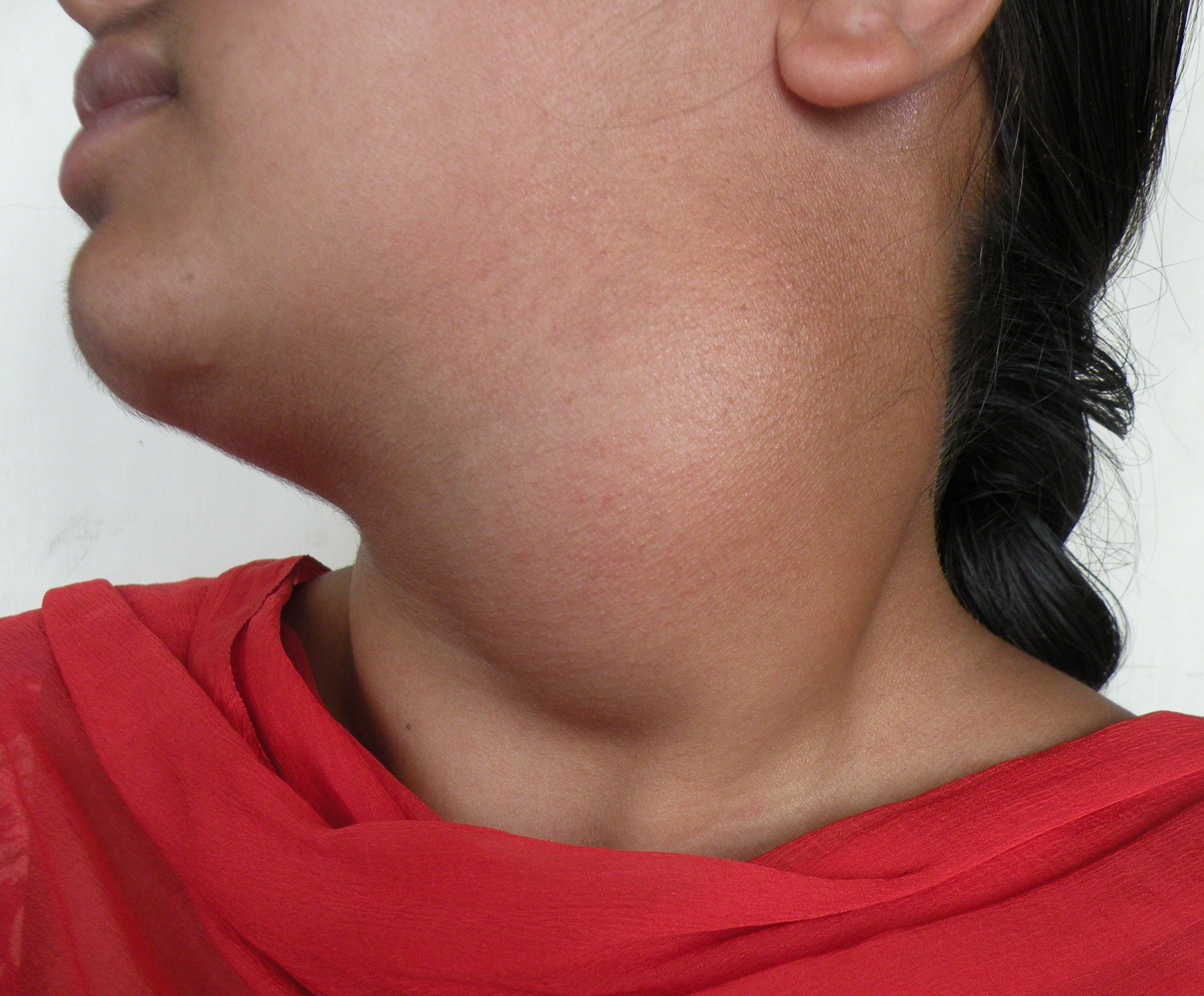
Thyroid lobectomy (right) for a deep seated nodule of right lobe. 55 years old female (c/o dr BL Bhardwaj). Hoarseness of voice persisted.
Prostatectomy (freyer) for a 120 grams huge prostate – easily shelled out – in a 60 years old. Haemostasis secured with sutures.
