Archive for the ‘Uncategorized’ Category
25.10.12
Re-exploration laparotomy for an obstructed gastric outlet, after laparotomy 3 weeks earlier by residents, in a young (20) male, with transection of duodenojejunal junction following trauma in a RTA. After the duodenal continuity was restored with a sutured anastomosis, a GJ had been added!. The patient continued to have more than 500 ml of bilious NG aspirate, and on removing the tube once, had some bilious leak from the abdominal wound, suggesting anastomotic leak, so the tube was put back in. At operation, the the DJ anastomosis was found to be strictured, and was dilated through an enterotomy with foley balloon and metal dilators, and the GJ was converted to RYGJ.
1.11.12
At lap chole, measured the dimensions of a Rouviere’s sulcus with the help of a feeding tube marked in centimeters, as a part of studying the anatomy of Rouviere’s sulcus as seen during laparoscopic cholecystectomy.

NCASICON at jalandhar – attended for two days : 8 and 9 september. Saw dr Chanjiv and others there.
13.9.12
Open prostatectomy for a huge (150 g) prostate who presented with hematuria. Postoperatively, developed clot retention, and had to be reopened.
Undescended testis in a 2 years old child- no testis found – anorchia.
20.9.12
Closure of ileostomy for a patient of Crohn’s disease who had presented with obstruction and residents excised a segment of ileum and had done an ileostomy. Patient on azathioprine by GE.
Revision of GJ into Roux loope GJ. An older patient who had presented in 2006 with GOO and had TVGJ performed. Now continued to have pain and vomiting intermittently. At operation, had stenosis of both limbs of GJ, which was revised into Roux-en-Y GJ. Did well thereafter.
21, 22 sept
attended SELSI hernia conference at Gwalior along with Dr Sushil Mittal and his team.
28.9.12
Abdominoplasty (assisted dr GP singh) for huge abdominal apron hanging down in a 45 years old female. Persisted with seromas for a long time.
3.10.12
dr Khushpreet’s case – laparoscopic excision of right ovarian cystic mass and left hydrosalpinx.
4.10.12
TEP repair of LIH in a 30 years old male.
11.10.12
TEP repair bilateral hernias 70 years old male. Had seromas both sides postop.
15.10.12
Mahesh Kumar, obese 50 male, admitted with severe pain in OPD. Very difficult lap chole due to obesity, gangrene of fundus of gallbladder, and thick walls making grasping impossible. Calots anatomy not clear. Ligated at lower part of gallbladder and then the suture used to retract the gallbladder further, then Calots defined. Took 4 hours.
22.10.12
Parotidectomy superficial for parotid duct stone and chronic sialadenitis, and stone not palpable intraorally.


9.8.12
Two clipless lap choles – the cystic duct and artery sealed with harmonic scalpel.
20.8.12
An abdominoplasty – assisted Dr GP Singh, the plastic surgeon. Dr Jagga’s patient – a young (25) female, who got a huge divarication of recti after delivery of her baby. The recti were plicated with 3 layers of vicryl.
30.8.12
Two TEP repairs for RIH – both easy and enjoyable.
3.9.12
Thyroid lobectomy (right) for a deep seated nodule of right lobe. 55 years old female (c/o dr BL Bhardwaj). Hoarseness of voice persisted.
Prostatectomy (freyer) for a 120 grams huge prostate – easily shelled out – in a 60 years old. Haemostasis secured with sutures.
Billroth II, diagnostic laparoscopy, and a series of difficult empyemas of gallbladder with fundus first dissection
Posted on: August 8, 2012
26.7.12 A huge duodenal perforation, closed by residents on 20.7.12, releaking. Young male (20 yrs old poor bhaiya). Explored to find a large perforation and posterior wall of duodenum stuck to pancreas. Mobilised with difficulty and duodenal stump closed. Billroth II type resection and GJ done. Duodenal stump blow out on 5th PO day.
2.8.12 diagnostic laparoscopy in a young (25 yrs old) male from Patran with chronic abdominal pain. Patient of dr Parmod Mittal. Endoscopy in 2011 showed some ulcerations in terminal ileum and ileocecal area – so took ATT for a year or so, but abdominal pain still persisting. At laparoscopy, had adhesions from previous open appy in RIF- these were divided. Running of all small bowel showed no stricture. In the left flank, was an omental band, adherent down in the pelvis; this was released.
5.8.12 four lap choles, 3 of which turned out be very difficult. The first one, an old blind lady (60 yrs old)- While dissecting with hook posteriorly, some bile leak was observed; changed gears to do the fundus first chole and the gallbladder ligated at its neck by no 1 vicryl loop. Drained bile tru the drain. ERCP next day revealed leak from CBD near CD-CBD junction. Stented – the drain dried up in 2 days. The second turned out to be an empyema unexpectedly. Was a younger patient (40 years female) and there was no sign of difficulty preoperatively. But it was a thickwalled empyema. Cut open to remove pus and stones. Ligated at neck. Third one was easy. The fourth againd turned out to be empyema. and same process was repeated.
Intestinal tuberculosis, glomus tumor, wrongly diagnosed cecal cancer, rectal prolapse, neglected torticollis and big incisional hernias.
Posted on: July 18, 2012
10.5.12
A 16 years old girl for laparoscopic appendectomy. Seen to have a terminal ileal mass at laparoscopy. The ileocecal segment was exteriorized laparoscopically, delivered out through a RLQ incision, and REEA was performed. Histopathology reported it to be tuberculosis of intestines.
17.5.12
A 50 years old policeman presented with recurrent discomfort in the left iliac fossa. He said he had visited all doctors around the area and all investigations done to no effect. At diagnostic laparoscopy, he was found to have a long adherent retrocecal appendix, the removal of which resolved the problem and the patient felt very grateful.
A clipless lap chole, the cystic duct sealed with harmonic scalpel in a 50 yr old female.
20.5.12
A small glomus tumor on left index finger excised under GA.
24.5.12
A young 30 years old prisoner, with history of appendix abscess drained in the unit on prisoner duty earlier, presented with a persistent mass in RIF. Was lingering on in the hospital for 4 months. FNAC of the mass showed only inflammatory cells. At operation, a densely adherent mass, adherent with terminal ileal loops, and cecum. All of it was excised and EEA done. Biopsy was reported as adenocarcinoma of cecum.
28.5 12
Dr Rama’s case: A 55 years old female, related to Dr Rama, reported with a thick walled gall bladder, with biliary stent in place at ERCP in Chandigarh for jaundice. Jaundice now relieved. With a thick walled gallbladder on CT and family history of cancer (cancer family syndrome), an open chole was done and cholecystectomy done with a 2 cm wedge of liver tissue. Biopsy however was nonmalignant luckily.
9.6.12
Held a clinical meeting in the ME cell of college. Delivered a talk on ‘The Symbol of medicine- the caduceus vs the staff of Aesculapius’.
14.6.12
A big incisional hernia repaired in a 60 years old female (relative of Fateh Singh, my neighbour). The hernia followed a resection of a big pelvic tumor (neurofibroma) at Patiala surgical center.
22.6.12
Laparotomy for perforation of ileum caused by the umbilical trocar inserted for diagnostic laparoscopy. Perforations closed and peritoneal cavity lavaged.
5.7.12
Five lap choles, 2 of which were clipless, done by harmonic sealing the cystic ducts.
9.7.12
A huge incisional hernia in a 55 years old female following open chole at Nawanshahar (c/o Surinder, Staff nurse in OT). A lot of terminal ileal loops and the whole of ileocecal junction and the right colon outside the abdomen. Difficult mesh repair.
Rectopexy (Wells) for complete rectal prolapse in a 60 years old man.
12.7.12
A bipolar sternocleidomastoid tenotomy for torticollis in a 16 years old female.
TEP repair of a right inguinal hernia in a 20 yr old male. Satisfactory.
An adult intussusception, Bhukki bolus obstruction, SMA syndrome, and a series of gallbladder empyemas.
Posted on: May 3, 2012
15.12.11 An adult intussusception in a 50 years old male. Ileocolic intussusception caused by a 3 cm tumor (histopath – fibrolipoma), REEA done. 

26.12.11
A difficult lap chole for empyema. 50 F. GB opened up to remove stones, and had to be removed piecemeal in 6 pieces.
Another difficult lap chole, 60 M, father of Ratan, house keeper with Dr DNB. Gangrenous gall bladder, separation of adhesions from duodenum led to a leak from duodenum. Converted to open operation to repair the damage.
29.12.11
An interesting laparotomy, a difficult one. 55F. Had a difficult surgery at Columbia Asia, reportedly for intestinal obstruction, due to a sigmoid colon cancer, and only biopsy and colostomy had been done. Presently, at operation, had a dense fixed mass in left paracolic gutter, fixed to anterior abdominal wall, was excised, and colostomy refashioned. Biopsy reported ovarian cancer. Later it transpired that she actually had a huge ovarian cystic mass removed at PSC last year and the biopsy that time was not malignant. The same mass had now recurred and involved the left colon.
2.1.12
Total thyroidectomy for a recurred goiter, subtotal done 15 years back.
10.1.12 TLH using ehicon enseal.
2.2.12
TEP was difficult, because the sac got torn.
Lap chole again for gangrenous gall bladder, duodenum got a tear, converted to open to repair.
6.2.12
An infected hydatid cyst of liver, adherent with abdominal wall. Laminated membranes removed piecemeal. A biliary communication ligated. Capitonage, and omentoplasty.
9.2.12
TEP, 60 M, difficult due to difficulty in creating the lateral space. Lap chole, again for empyema. GB removed piecemeal.
16.2.12 Difficult lap chole, short thick cystic duct, ligated with Roeder knot.
23.2.12 TEP, 25m, easy.
1.3.12 TEP , 25 m, difficult again due to problems in creating the lateral space.
19.3.12 A wrong diagnosis of an ovarian cyst was made (on US findings) in a 35 y o female; turned out to be a big chronic pelvic abscess with thick adhesions, and a left TO mass; the mass was excised, and the pus drained. Recovery from GA delayed, had to kept in ICU on ventilatory support for two days.
22.3.12
Lap chole, again for empyema, the stone at neck removed first, and GB removed piecemeal.
24.3.12 Lap chole, converted to open, because of adhesions with duodenum, subtotal chole only possible.
26.3.12 Lap chole, 130 kg obese 35 female, wife of Sahib Singh Cheema from kheri, had to be converted for huge empyema. A small PUH repaired at the same time, on patient’s request; got into trouble later because of wound infection, which did not clear with dressings till 3 may, when it was explored again under GA and a deeper abscess drained.
29.3.12 DJ (Duodenojejunostomy), stapled, for SMA syndrome, in a young (15 years old ) girl, thin and emaciated. Recovered well.
2.4.12 A very difficult lap chole, again for empyema, 50 F. Wall of gall bladder more than 1 cm thick, had to be bisected for the grasper to hold. Stones removed, and gallbladder had to be cut into 6 pieces to remove from the epigastric port. Took nearly 3 hours. Ileus for 2 days.4.4.12
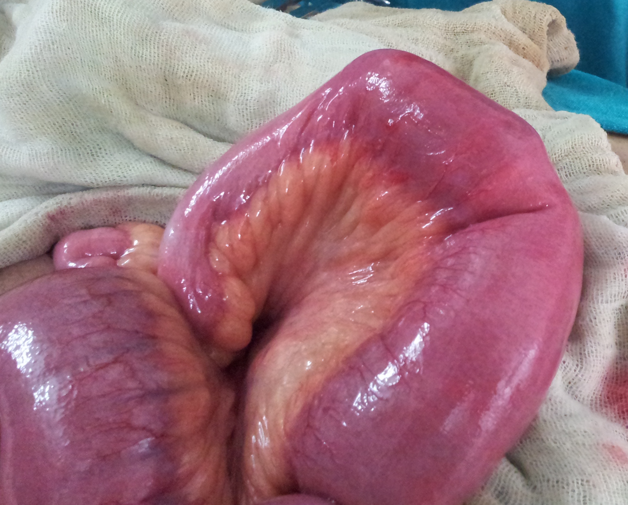
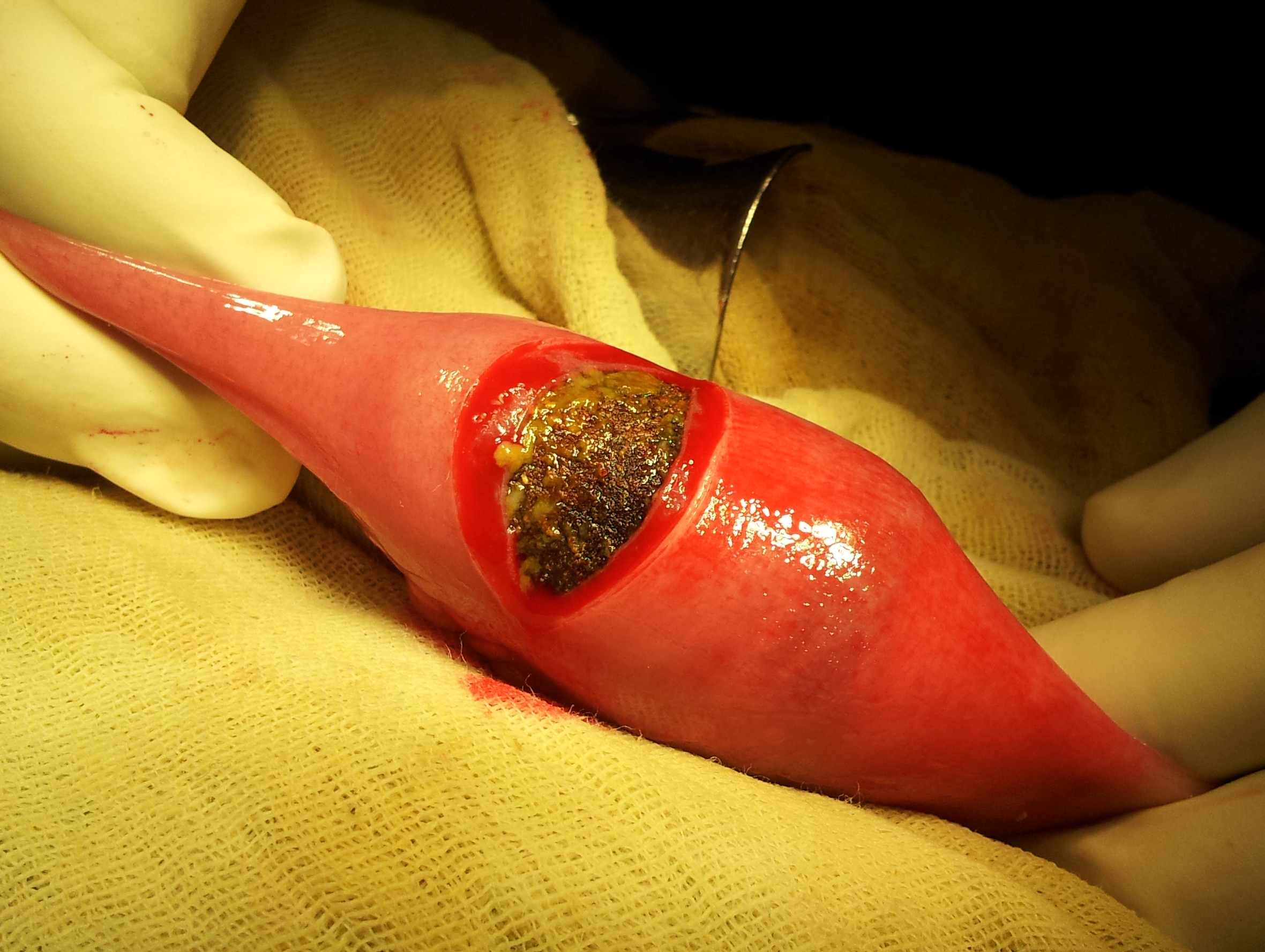
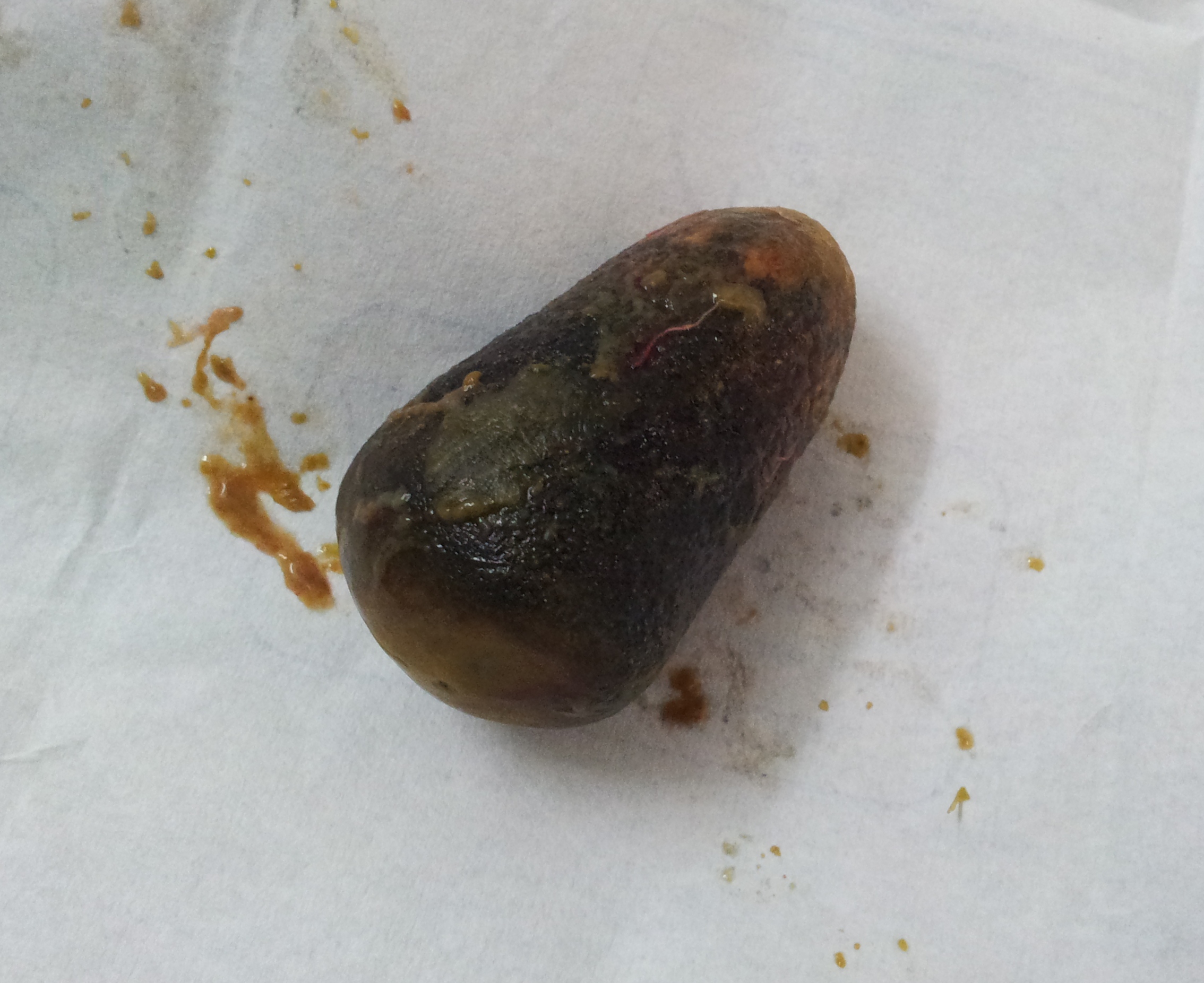
A unique cause of intestinal obstruction. A 30 year old young man, addicted to poppy husk (ver: bhukki), with small bowel obstruction, found to have a bolus of inspissated poppy husk ( bhukki) in the ileum, removed through an enterotomy.
9.4.12
Lap chole, 50 year old, c/o Surinder, staff nurse in OT. Thick-walled empyema, had to be converted due to dense fibrosis in Calot’s triangle. Stones removed after opening gallbladder and the GB ligated at its neck.
12.4.12 Another empyema of gallbladder, could be done laparoscopically.
26.4.12 Lap chole in a child 7 years of age. Difficult. Turned out to be a thick walled gall bladder with a walled off perforation at the fundus, discharging pus after removing adhesions. Dark sludge in the gallbladder. GB too thick to come out of the 10 mm port, had to be removed piecemeal.
Lap chole, 50 f, most difficult again, had to be converted due to dense adhesions in the triangle of Calot. Fundus first dissection deemed safe. Neck suture ligated. A small perforation in duodenum closed.
30.4.12 Lap chole, converted again, for thick-walled empyema. GB opened, stones removed, and the pedicle ligated. Small ooze of bile observed middle of liver bed from a small calibre cholecystohepatic duct, ligated. Still leaked bile for 3 days postoperatively, then dried up.
Lap nephrectomy, 30 M, for NFK (PUJ) right kidney, pedicle dissection deemed too dangerous, so converted. At open operation too, the pedicle could not be separately defined.
1.8.11
An obese 50 F with a big right thyroid swelling; at operation, the big cystic swelling removed had actually no connection with the thyroid. Biopsy reported as colloid goitre!!
4.8.11 A big swelling in the floor of mouth as well as under it – diagnosed as big ranula; at operation turned out to be a big dermoid cyst!!
6.8.11 Went ot kurukshetra to attend MACT; saw dr subhash too.
17.8.11 A difficult lap chole for thick walled gallbladder and a big impacted stone in neck, making grasp difficult. The stone extracted first, then grasp was possible.
28.8.11 A big ovarian cyst removed laparoscopically- biopsy was reported as endoetrioma!
1.9.11 A satisfactory TEP at last. Small indirect sac and a lipoma of cord.
5.9.11 TEP converted to open because space could not be created after the first trocar went intraperitoneal.
6.9.11 Laparoscopic gonadectomy for testicular feminization syndrome. Interesting case.
12.9.11 A neat TEP repair of a small indirect hernia.
19.9.11 Another easy and quick TEP repair for bilateral hernias; heavy mesh on one side and lightweight mesh on the other side.
20-24 september: Attended the ICS conference at Srinagar.
9.10.11 A combined lap chole and lap excision of ovarian cysts bilateral – difficult chocolate cysts on both sides. Took more than 2 hours.
13.10.11 A displaced cu-T enmeshed in omentum removed laparoscopically.
17.10.11 An unusual GOO – GJ done. 55M with GOO. CT showed mass outside stomach. At operation, a matted mass wedged in D2 and head of pancreas.To be planned later a possible whipple.
23.10.11 A sad case of a young (30) female, with advanced rectal cancer causing intestinal obstruction. At operation, diffuse carcinomatosis in whole of peritoneal cavity and a big fixed recal tumour. Even small bowel loops matted in the lower abdomen. One upper loop brought out as ileostomy.
6.11.11 Another strange case diagnosed as pleomorphic adenoma of parotid on FNAC and CT. At operation, superficial parotidectomy was done but the two cystic masses present in front of and adherent with the parotid tissue and removed with parotid turned out to be tuberculous on biopsy.
7.11.11 Cone excision of breast ducts (Hadfield). 50F, relative of OT staff nurse Surinder. Had two operations for nipple discharge earlier.
10.11.11 A planned lap chole in a 60 years old man with pain abdomen and US showing gallstones, postponed on finding an umbilical nodues from which an FNAC was done. This showed adenocarcinoma.
30.11.11 A planned femoral hernia repair in a young man of 20 years was postponed on finding his BP to be 210/110. To be investigated for hypertension first.
1.12.11 A laparotomy for gastric cancer turned out to be futile on finding the whole stomach fixed with no available area for a GJ. Omentum full of deposits.
16.5.11
SCC foot excised in a 60 yr old male. Biopsy reported melanoma. To start with chemo later.
19.5.11
Laparotomy for intestinal obstruction 55 years old male with history of right hemicolectomy for cancer right colon; gut loops stuck to a hard mass (fixed and unresectable) midileum. Only a side to side jejunoileal anastomosis possible for to relieve obstruction.
26.5.11
Huge incisional hernia (following a Hartmann’s and later its reversal for cancer colon), 55 years old male from Mukatsar, repaired with two large meshes.
28.5.11
A clinical meeting held in medical education cell, Dr Kundal lecturing on PBF.
1.6.11 to 7.7.11 summer vacation.
2.6.11
Lap chole (c/o Dr Sharma from Dhuri), difficult but had to be converted to open due to failure of electric supply to OT!
7.6.11
Went to Bathinda medical college with Dr Jagga. Saw Dr Ravi, Dr Kiran and Dr Dhaliwal there.
2.7.11
Went to Bathinda again for Ginu’s practical examinations starting today.
11.7.11
Three lap choles
14.7.11
Three lap choles again.
18.7.11
Hepaticojejunostomy Roux-en-Y for a suspected choledochal cyst (reported on US and MR). Hamidan 55 years old with h/o open chole and CBDE for jaundice and gallstones at KMH by Dr Khurana 25 years back. Now had pain upper abdomen. LFTs mildly deranged and CBD reported dilated 2 cm, with small stones in it. Refused ERCP and wanted an open operation. At operation CBD found to be dilated 1.5 cm with some concretions which were flushed out. A Roux-en-Y hepaticojejunostomy was carried out.
25.7.11
Four lap choles.
9.4.11
After a long time, a clinical meeting was held in the college ME cell (medical educaition cell). I presented NMS (neuroleptic malignant syndrome) there.
11.4.11
A most difficult lap chole in a 55 M, gallbladder ligated at neck. POD 2 – bili 6, raised enzymes too. Sent to PGI for MRCP and nuclear scan, which were normal!. Remained remarkably fit and normal clinically. Reason for the raised bilirubin and enzymes??
12.4.11.
TLH.
19.4.11
A very difficult lap chole again in a young female. Thick walled empyema. Gallbladder cut into 5 pieces to remove.
30.4.11
Second clinical meeting. Dr Mohi presented a germ cell tumour arising from abdominal wall!!.
2.5.11
Another very difficult lap chole. Very dense adhesions, their separation resulted in stripping of serosa of duodenum. Converted. A tear in duodenal mucosa had to be sutured.
4.5.11
Nephrectomy (left) for a huge pyonephrosis in 60 y o male. Dense vascular adhesions with left colon and spleen and pancreas.
5.5.11
TEP for RIH. Previous left inguinal open repair already done. Peritoneum breached and the sac too got opened up!.
ASI NC conference at Simla attended from 6 to 8 may.
12.5.11
3 lap choles and a TEP RIH repair.
4.4.11
A very neat, quick and easy lap chole.
Near total thyroidectomy for toxic multinodular goitre, failed on medical treatment: relapsed after one and a half year of treatment. No more than a gram of tissue had to be left surrounding the right recurrent laryngeal nerve.
