Archive for the ‘operations’ Category
VVF repair and summer vacation
Posted on: June 20, 2010
18.5.10
50F with a right thigh swelling, twice recurred and now causing excruciating pain. Previous biopsy fibrous histiocytoma. Now again excised, but with wide (2 cm) margins. Skin closed under tension. Wound gaped after 7 days.
21.5.10
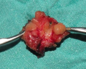
1. Young female (16 Kammo’s daughter). Two fibroadenomas apparently looking like one, excised under GA.
2. a big preauricular swelling (possibly dermoid) on right side in a 10 year old female child, excised under GA.
25.5.10
1. 50 year old obese female with varicose veins right leg. Trendelenburg + stripping + multiple phlebectomies.
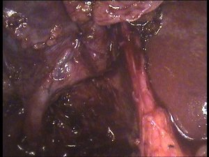
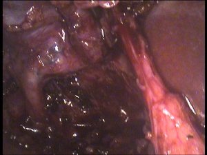
2. VVF repair in a poor 60 year old lady who had the fistula for 15 years following a hysterectomy. Abdominal repair after splitting the bladder anteriorly. Fistula rather low down and between the two ureteric orifices which were identified and protected.
28.5.10
3 lap choles one after the other. The first two were easy. The third, Alka Rani (c/o Dr Suman from Phagwara), 50 F, hypertensive and diabetic, had a thick walled empyema. 200 ml thick pus aspirated. Difficult dissection in Calot’s triangle. Had fever postop for some days. Then gradually settled.
1.6.10 to 7.7.10
Summer vacation
6.6.10PMT test for binu and ginu at faridkot. binu got 17 rank and ginu 531.
MRM left breast for a big tumour (looked like cystosarcoma phyllodes, bosselated). Just one or two lymph nodes in the axilla.
19.6.10
Went to delhi with binu and Dr Jagga for binu’s CBSE counseling. Binu selected Patiala medical college. Returned late in the evening to Patiala.
20.4.10
Lap chole in a very obese (>120 kg) 35 years old female. Difficult due to fat in Calot’s triangle. Had to be kept in anaesthesia recovery for a very long time due to respiratory problems.
23.4.10
Varicose veins leg 35 M – Trendelenburgh, stripping to knee and multiple phlebectomies.
27.4.10
Thyroid lobectomy for recurred thyroid cyst after aspiration. 50F.
4.5.10
Extended right hemicolectomy 55 M from Dhanaula, with h/o anaemia, occult blood in stools positive and CT showing a mass in right colon. At operation, the cut section of specimen showed no real tumour inside but instead 3 strictures and multiple lymph nodes in mesocolon.Biopsy reported as eosinophilic colitis!!.

7.5.10
60M with h/o severe pain after every feed, CT showing dilated D1 and D2 and giving a diagnosis of SMA syndrome. Laparotomy – lot of ascitic fluid, missed on imaging!!. DJ flexure tightly fixed and scarred with nothing passing beyond into jejunum. No real tumour palpable. FNAC taken from dense retroperitoneal scar. GJ done. Cytology of ascitic fluid and FNAC came out to be negative for malignancy.!

11.5.10
TEP repair for bilateral inguinal herniae. 35M from Samana (dr Ashok Galib’s case)
12.5.10
Laparoscopic nephrectomy right non-functioning kidney.
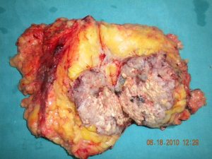
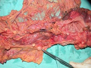
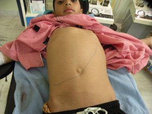
Nephrectomy for RCC
Posted on: April 18, 2010
16.4.10
Nephrectomy for renal cell carcinoma lower pole right kidney. 35 M. BJP worker.Remains well on follow-up as of now (2013 april)

Some strange times
Posted on: April 11, 2010
- In: operations
- 3 Comments
26.3.10
Swelling left upper arm excised under GA. ?STS
28..3.10
Lap chole for acute cholecystitis, supposed to be routine. 35 F. US – polyp or sludge?
Actually a mass in fundus palpable at lap chole. Aspiration yielded dark blood!
Tense oedematous cholecystitis. Routine lap chole done. Biopsy adenocarcinoma.
30.3.10
Pilonidal sinus excised with primary closure. Did well. No recurrence.
31.3.10
Abdominal wall right flank incisional hernia repaired with mesh. 35F Sikh lady from Pakistan!!, with a history of RPM laparotomy (for what ?) with right flank drain in Pakistan. Hernia had developed at the drain site.
2.4.10
35M with previous h/o admission for appendix mass 3 months back. Hard mass still palpable. Ba enema reporting a filling defect. At right hemicolectomy, mass found to be posterior to cecum, but fixed to cecem and immobile. Could not be completely removed. Biopsy – inflammatory cells in one area behind the caecum – giant cells and fibroblasts. ?diagnosis.?bony. To be reassessed later.
5.4.10
To Simla to attend a second review committee to review the questions of MS/MD examination conducted by Simla university. Himachal high court had ordered to review the questions after students challenged some questions. The first review committee had only local faculty, so court asked for additional members from outside Himachal.
6.4.10
Thick walled empyema in 60 M. Lap chole.
7.4.10
Huge hydatid cyst superior half of right lobe of liver. 14 years old boy. Membranes removed intact and in toto. Huge cavity had to be capitonnaged and introflexed. Omentum not developed enough to bring into it. 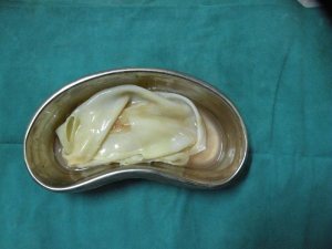
9.4.10
Had to atttend to summons from SP(D) (superintendent of police, detective) enquiring into complaint by relatives of a patient Manjit Kaur who had died of septic complications after left hemicolectomy for obstructing large splenic flexure adenocarcinoma; the patient had a leak and was reexplored 3 days later. Patient’s mother alleged that her kidneys had been removed!. What a change of times!
10.4.10
Imperforate hymen in 14 years old girl incised and dark blood (200 ml) drained.
11.4.10
TAH in a 12 years old girl!!. Mentally retarded with cerebral palsy and urinary and fecal incontinence. Mother’s insistence for the operation.
26.2.10
Lap chole in a 30 years old male (a surgeon himself, assistant professor, GSMedical college). Turned out to have a rather prominent accessory bile duct arising from the middle of the liver bed and draining directly into the gall bladder. After much deliberation and hesitation, this was doubly clipped and divided. Concerned at the prominent size of this duct though. Postoperative OK so far.

Right parotidectomy for chronic sialadenitis, causing pain and swelling during eating for last 10 years. 30 years old male. At operation, small pockets of pus in the parotid tissue. Dense fibrosis at places, making dissection of facial nerve branches difficult. Nonetheless all branches displayed and protected. Biopsy – chronic parotitis.
2.3.10
Easy male gall bladder in a 60 years old patient! Pleasant surprise.
Psoas abscess drained in patient with history of nepherectomy for RCC, followed by radiotherapy. Radiation induced myonecrosis?
3.2.10
TEP for bilateral inguinal herniae. Initial entry into preperitoneal space too superficial, nearly avusling the inferior epigastric vessels, which were coagulated eventually because of the continous bleeding.
17.3.10
Sacrocolpopexy for vault prolapse. Mesh strip buried under the peritoneum.
Re-exploration of rectopexy, etc
Posted on: January 28, 2010
30.12.09
Rectopexy done 10 days back re-explored for adhesive intestinal obstruction.Adhesions in pelvis lysed.
5.1.10
Infiltrating type of huge lipoma thigh excised. 55F patient of dr Mrs Hans
12.1.10
3 lap choles.
18.1.10
Big perinephric abscess drained. Dr Jagga’s case.
22.1.10 Nephrectomy for RCC right kidney. 65 M.

28 and 29 november
Went to Kasauli with Dr Hardip Singh Mann and joined other classfellows of the 75GOMCO batch for a get-together. Stayed overnight and came back next day. Met old friends – dr Dharam Pal, Dr Tarsem Rattu, Dr Ravi Dawra, Dr Brij Kishore, Dr Bhucho (Keshav Garg), Dr Zora Singh, Dr Sohan Singh, Dr Gursharan Malhi, Dr Surinder Gupta, Dr Harjit Singh (braather), and many more.
30.11.09
Wide local excision (a near mastectomy) of an indeterminate mass despite 2 FNACs and mammography in an 85 year old female (an ex health worker – ANM). HPE- IDC. Started on tamoxifen.
A potential disaster averted just in time – lap chole in a 65 year old male, very difficult procedure. Liver firmly stuck to abdominal wall and absolutely immobile. Thick walled gall bladder with a very difficult grasp. A very easily dissected CHD, misidentified as CD, was clipped and about to be divided, when better sense prevailed, clips removed and a further dissection revealed a very short and thick cystic duct which was ligated with extracoporeally tied vicryl. Stormy postoperative period with abdominal distension and vomiting, the NG tube poured greenish output for a couple of days and then everything settled!! Could never tell what happened!!. A postoperative US scan and LFTs were normal.
1.12.09
Units of surgery department reshuffled after retirement of Dr Karam Singh. Mine will be 4the now, ward 3.
4.12.09
Microdochectomy for a single duct discharge from left nipple. 30 F, a principal of a school. Biopsy showed a well-formed papilloma.
9.12.09
Seminar on early detection of breast cancer.
11.12.09
A recurred swelling left forearm near the lower end of ulna. Presumably lipoma, turned out to be a swelling arising out of the tendon sheath of the extensor of the little finger, the ulnar artery pulsating close by. Excised under GA, preserving the tendon.
20.11.09
Laparoscopic excision of a big paratubal cyst (6 cm). Rather than the cyst alone, the whole mass of the cyst, tube and ovary was excised at the request of the attending gynaecologist. The other tube also coagulated with bipolar diathermy and cut (patient’s demand).
23.11.09
Carcinoid ileum – Right hemicolectomy for a presumed cancer caecum. A big mass palpable in RLQ of abdomen in a 20 years old female!. Getting obstructed with solid feeds, so kept on liquids for some days. CT showed mass in the caecum and ascending colon. Cut section showed mass in wall of terminal ileum rather than the lumen of caecum. Turned out to be carcinoid on histopathology and immunohistochemistry.

5.11.09
1. Big hydatid cyst right lobe of liver pressing on gall bladder and bile ducts, leading to elevated bilirubin levels. Laminated membrane excised in toto in one piece, and cavity partly obliterated (capitonage) and then filled with omentum. No drain.

2. Laparotomy for a mass lower abdomen and a vesicoenteric fistula, presumed to be a desmoid of abdominal wall. 17 M emaciated with history of blunt abdominal trauma, followed by history of urine contamination with faeces. At laparotomy, dense mass in lower abdomen with underlying loops of bowel adherent with it. Opened accidentally (and repaired) the transverse colon loop here while opening the abdomen. Painstaking dissection identified the proximal small bowel loops but the distal ileum, converted into a mass which was adherent to the bladder, had to be excised and EEA done. The bladder fistula was a small one and was closed.
8.11.09
30M taken up for appendicectomy thru small RLQ incision had to be converted into upper midline laparotomy on finding turbulent fluid in the peritoneal cavity. A DU perforation closed.
12.11.09
Two difficult lap choles:
1. Tripat Chand 72 years old male from Barnala (referred by Dr Surinder Garg and also known to Dr Avinash Gupta, AP orthopedics). Had a mass covered with omentum as if it was covering the gall bladder but on some dissection, a non-adherent gall bladder could be visualised behind this mass. Routine cholecystectomy was done with some difficulty due to the mass coming in the way. The mass (possibly with transverse colon or distal stomach) to be investigated later with CT and other investigations.
2. Post pancreatitis (gallstone) lap chole. 35F. Thick walled cystic duct, endlooped and ligated.
16.11.09
Laparoscopic ureterostomy (Dr Sukhpreet’s case) for a presumed stricture or radiolucent stone right midureter in a 30 years old female with pain and right hydronephrosis and hydroureter ureter above the stricture. Transperitoneal laparoscopic approach easily identified the bulge in the right ureter just below the caecum and over the pelvic brim. Peritoneum over it opened and ureter opened longitudinally over it with hook cautery to reveal the curled up stent in it but no stone. The thick wall of the stricture was further opened up and the stent pushed up into the renal pelvis. Uterer closed over it with 3-0 vicryl and the peritoneum too was closed over it.
17.11.09
A most difficult lap chole. 65 F obese patient. Tense empyema aspirated to begin with. The neck of gall bladder found to be very firm as if it contained a hard impacted stone in it. This neck was opened up to find a very thick walled abscess here. Dissecting below this could have been dangerous, so fundus first dissection was done upto this point and a subtotal chole was done. Three big stones removed from within the opened up gall bladder. These were broken into pieces and removed piecemeal. Then the gall bladder walls were also cut into several pieces and removed . Drained.
18.11.09
A difficult TAHBSOP. 40 F with previous unsuccessful attempt at Sirhind. The problem was the big fibroid with posterior wall of uterus. This was stuck in the pouch of Douglas, and pressing upon the displacing the right ureter.
25.9.09
3 difficult cases –
1. TAHSOP
2. Appy lap : 30 M handicapped (RLLPPP) with h/o burst mass appx 6 months back. Tip of appendix still badly stuck retroperitoneally.
3. Laparotomy for neglected intestinal obstruction. Poor young (30) female, with previous 2 LSCS. At op, gangrenous loop of jejunum (thick omental band in pelvis from previous CSs) resected and EEA done.
27.9.09
Lap chole in 55 M (brother of Dr Ravinder Singh ex-prof orthopedics), difficult. Thick walled gall bladder and the thick short CD end-looped.
5.10.09
Lap chole converted to open. 70F with previous ERCP and ES for CBE stones. Dense adhesions with stomach. Calot’s triangle not clear even at open. Big stone impacted in neck removed and the neck ligated.
7.10.09
Laparoscopy for right ectopic tubal pregnancy which had already aborted thru fimbria. Thus tube could be saved after securing haemostasis from the fimbrial margins.
17.10.09
diwali
19.10.09
seminar on small bowel tumours.
20.10.09
Tried removal of a big chocolate cyst left ovary (dr jagbir’s patient) but had to convert due to the big size of the cyst (reaching upto above the umbilicus) and adhesions due to previous surgeries (CS and TAH). Even at open, was a tedious job.
22.10.09
Lap chole, day case, 52 F with acute cholecystitis, and thick walled gall bladder.
Nephrectomy (L): 30F with non-functioning kidney from long-standing PUJO. Calcified hydronephrosis. Retrospectively, could well have been done laparosocopically.


