Author Archive
Omentocele stuck to testis, a torticollis, and a bile leak in a difficult LC
Posted on: April 27, 2024
27.9.23
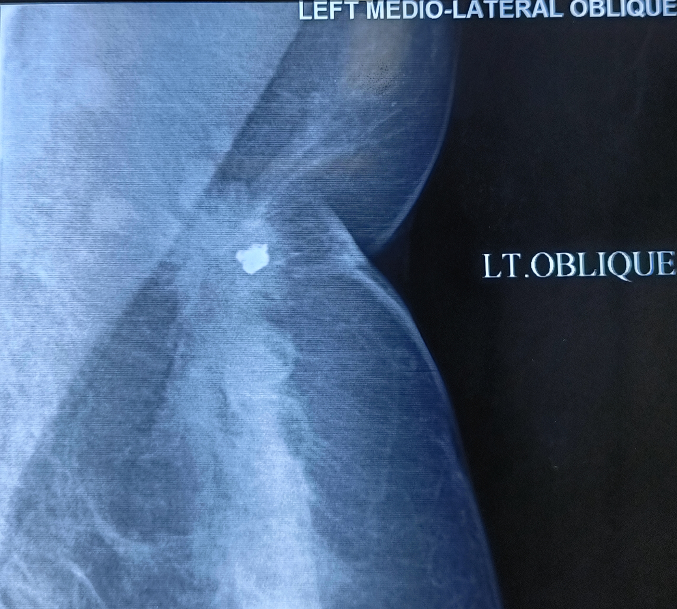
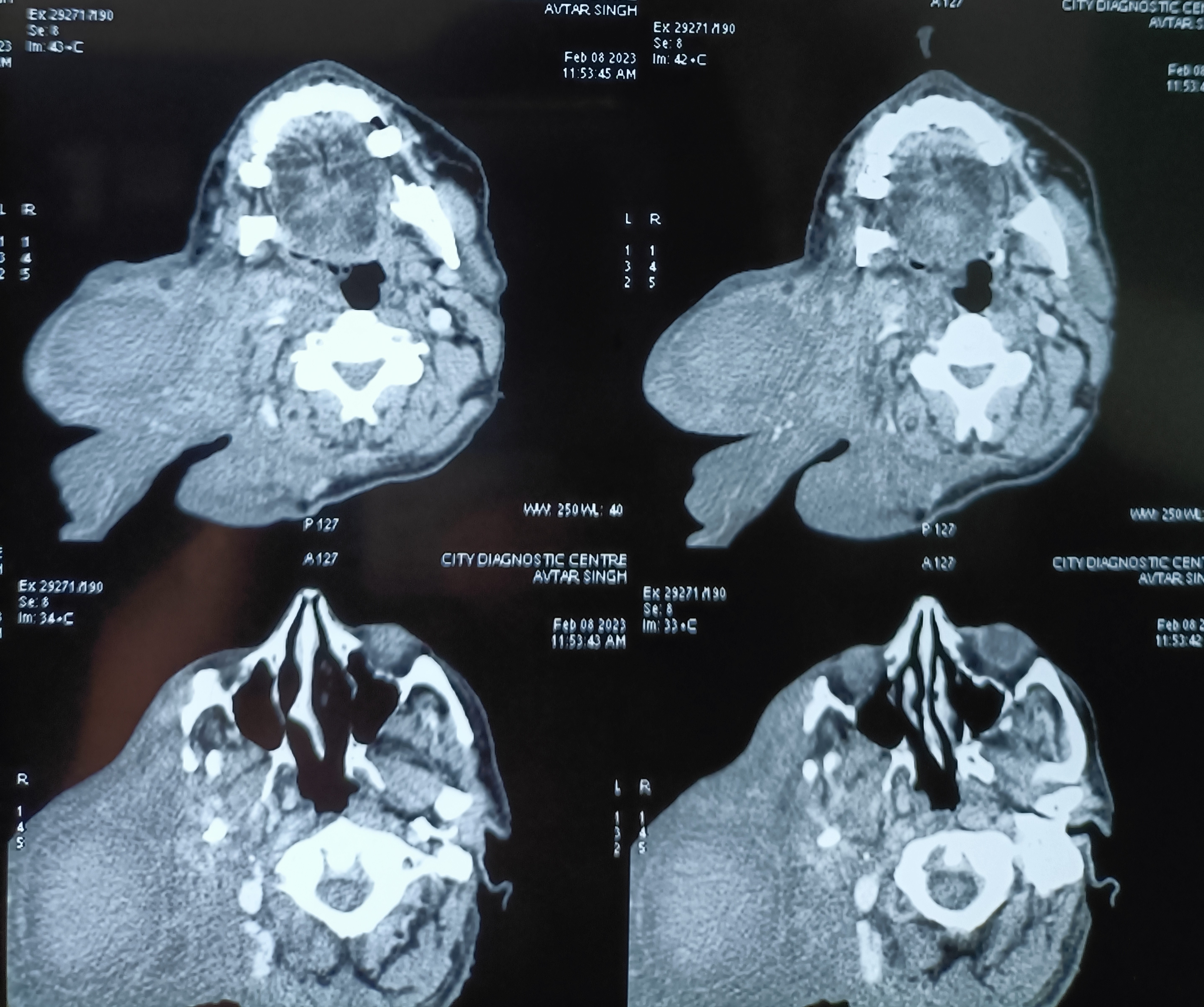
A 28 years old male patient (related to an employee of saket hostpital) presented with torticollis since childhood, with severe deformity of neck and face. The neck X-ray revealed scoliosis, making intubation for anesthesia difficult.
A bipolar sternocleidomastoid tenotomy was performed, and a cervical collar given.
21.10.23
During dissection for a big right inguinal hernia, it was found that the big omentocele was adherent to the testis and appeared to be going into the testis. And indeed, the capsule of the testis had to be incised to deliver the omentum from within the testis.


28.11.23
During a difficult dissection in a lap chole, cautery dissection lower down on the Hartmann pouch resulted in a tiny perforation which resulted in a bile leak for a week.
19.6.2023
RIH indirect hernia repaired in an obese 50-year-old male patient. The indirect sac was ligated and excised as usual. Also had a big lipoma in the canal quite free from the cord – unusual. This was excised too.
20.6.23
Urethral caruncle in an elderly 65 years old female had caused urinary obstruction and stricture of the distal urethra. Was catheterized after excision of the caruncle and urethral dilatation.
30.6.23
Mesh removal in a 67 years old male (brother of Cheema Sahib Singh) following meshalgia and a chronic sinus developing after a hernia repair in 2022. The mesh was removed with difficulty. The wound developed a hematoma and infection for which the nonhealing wound was laid open on 16.7.23. After this, the wound gradually healed.
9.7.23
A difficult lap chole in a diabetic and hypertensive 55 years old male took more than 2 hours but was eventually successful when the gall bladder was emptied of the big stones first, and the big Hartmann pouch was lifted high up to reveal a fibrotic calot’s triangle. Dissection starting higher up from the left could achieve a plane of dissection behind the neck of gallbladder, at which level it was ligated with vicryl. Earlier attempts at dissecting from the right on the H. pouch were not successful.
Craniectomy bones in the thigh forgotten for more than 10 years, and an abandoned APR for GIST of the rectum
Posted on: September 20, 2023
19.5.23
A 76 years old rural lady had undergone brain aneurysm clipping at PGI in 2010, when the craniectomy bones were kept in a subcutaneous pocket in the thigh. She never reported back there for removal of these bones from the thigh, and forgot about them. Now after 13 years she presented at TNH with a large palpable swelling over the thigh with a discharging sinus. The bones were removed and the wound which had a large collection of serosanguinous fluid cleaned and debrided of several areas of inflammatory granulations, drained and sutured.


27.5.23
A large paraumbilical hernia (supraumbilical) repaired with mesh in a 62 years old hypertensive, diabetic and obese lady (w/o Ranjit S Bhullar). The incarcerated omentum looked like gut loops, eventually dissected out completely and excised. Drain removed after 5 days, but the wound continued to discharge and took exactly a month (till 27 June) to dry up.
An attempted APR (case of Dr Sukhpreet) was abandoned in favour of palliative sigmoid loop colostomy in a 60 years old male patient, who had a big mass in the rectum diagnosed as GIST (spindle cell type) on biopsy. At exploration, the mass was found to be immobile, with vascular angry-looking surface; and the resection was abandoned. The sigmoid colon was found to be large and fixed in the right side; so had to be mobilised and and a loop colostomy made in the RIF.


Chronic venous leg ulcer, persisting despite multiple interventions; and a breast cancer incorrectly diagnosed
Posted on: August 20, 2023
5.5.23
A 45 years old patient presented with a history of suffering for 5 years from varicose veins and a non-healing ulcer in the right lower leg. Had undergone surgery for varicose veins twice in RHP, first in 2018 and then again in 2020. A third surgery was done in a private hospital in 2023. On examination the ulcer and varicose veins were still present. Was advised a repeat venous doppler ultrasound study, and compression dressings/stockings in the meantime. He never reported back. However, the two prescriptions he carried were the following.


4.5.23
A wide local excision of a breast lump was carried out in a 45 years old female (Dr Jagga’s case), presuming the the lump to be not malignant because the FNAC had not reported carcinoma, but only atypical ductal hyperplasia. However, at operation a clear skin dimple was noted in the left upper quadrant, strongly suggesting cancer (this clinical sign was perhaps not noted by the clinician). The mammography report was BIRADS IV/V category (highly suspicious).


The biopsy report, as expected, was IDC. An MRM was done on 25.5.23.
2.5.23
A 7 years old male child admitted with acute abdominal pain showed classic signs of target sign/doughnut sign/pseudo-kidney signs on ultrasound and a mass effect in right iliac fossa on CT. However, the child’s condition improved rapidly and in a few hours he became clinically better and pain free, suggesting spontaneous reduction of the intussusception. Recovered completely on conservative treatment and was not operated.


3.5.23
An elderly man (70 years old), a Nihang, had been having an inguinal hernia for nearly 20 years. This had now grown hugely, extending down into the scrotum, with abdominal X-ray showing signs of intestinal obstruction. However, he was adamant at not having surgery.

12.2.23
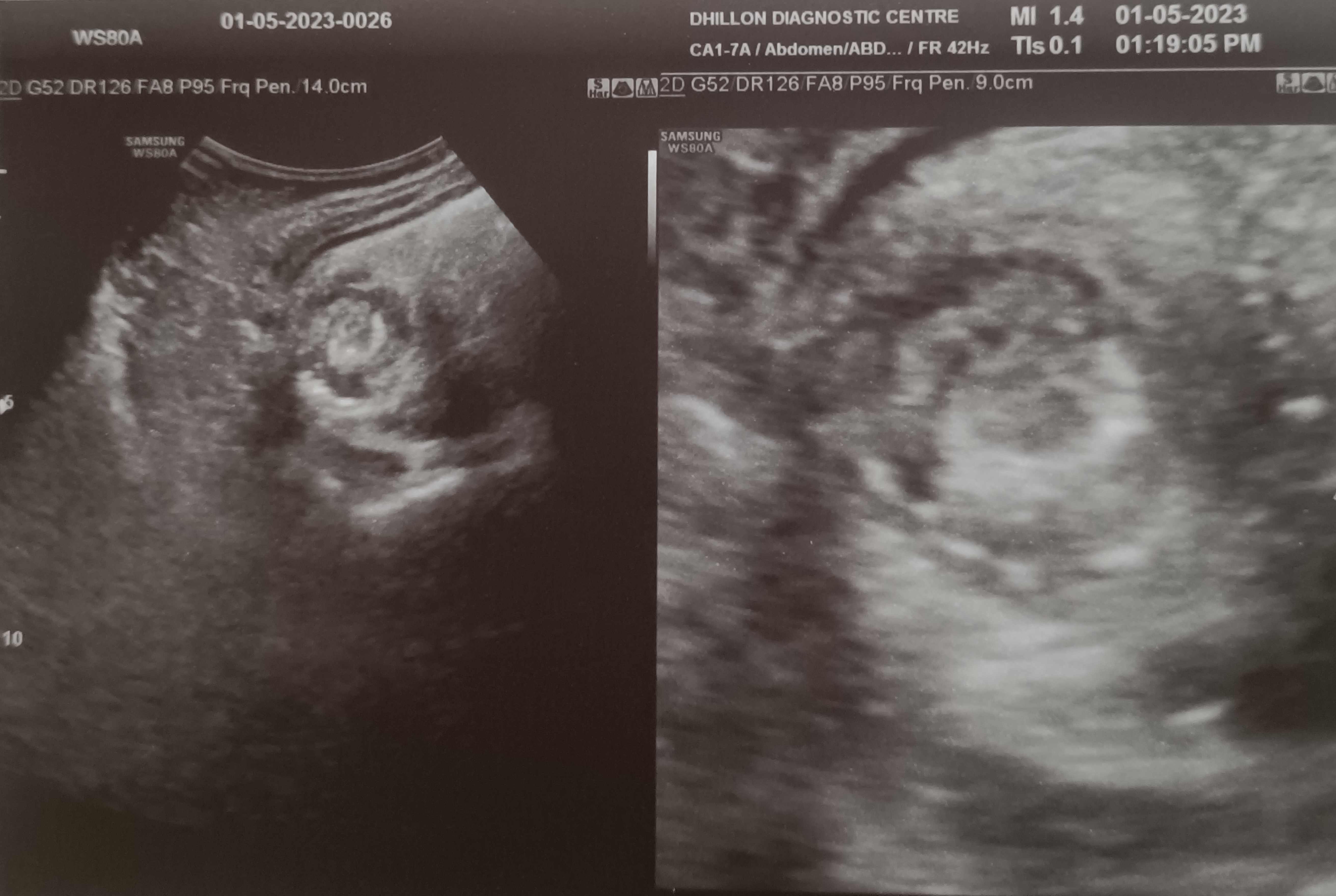
A 35 years old female (from Sunam) presented with a chronic left parotid swelling. An ultrasound scan reported a heterogenous mass in the left parotid gland, and a CT scan showed a 22×42 mm sized mass in the superficial lobe; the FNAC suggested chronic reactive lymphoid hyperplasia. At superficial parotidectomy, the superficial lobe was found to be diffusely enlarged (no discrete mass with discrete margins), and was removed. Biopsy reported reactive lymphoid hyperplasia.


Feb, 2023
An old man, nearly 70 years of age, presented in OPD with huge plexiform neurofibromatosis of the face and back, with a history going back to more than 50 years. He was now distressed particularly by the big facial lesion and wanted excision.

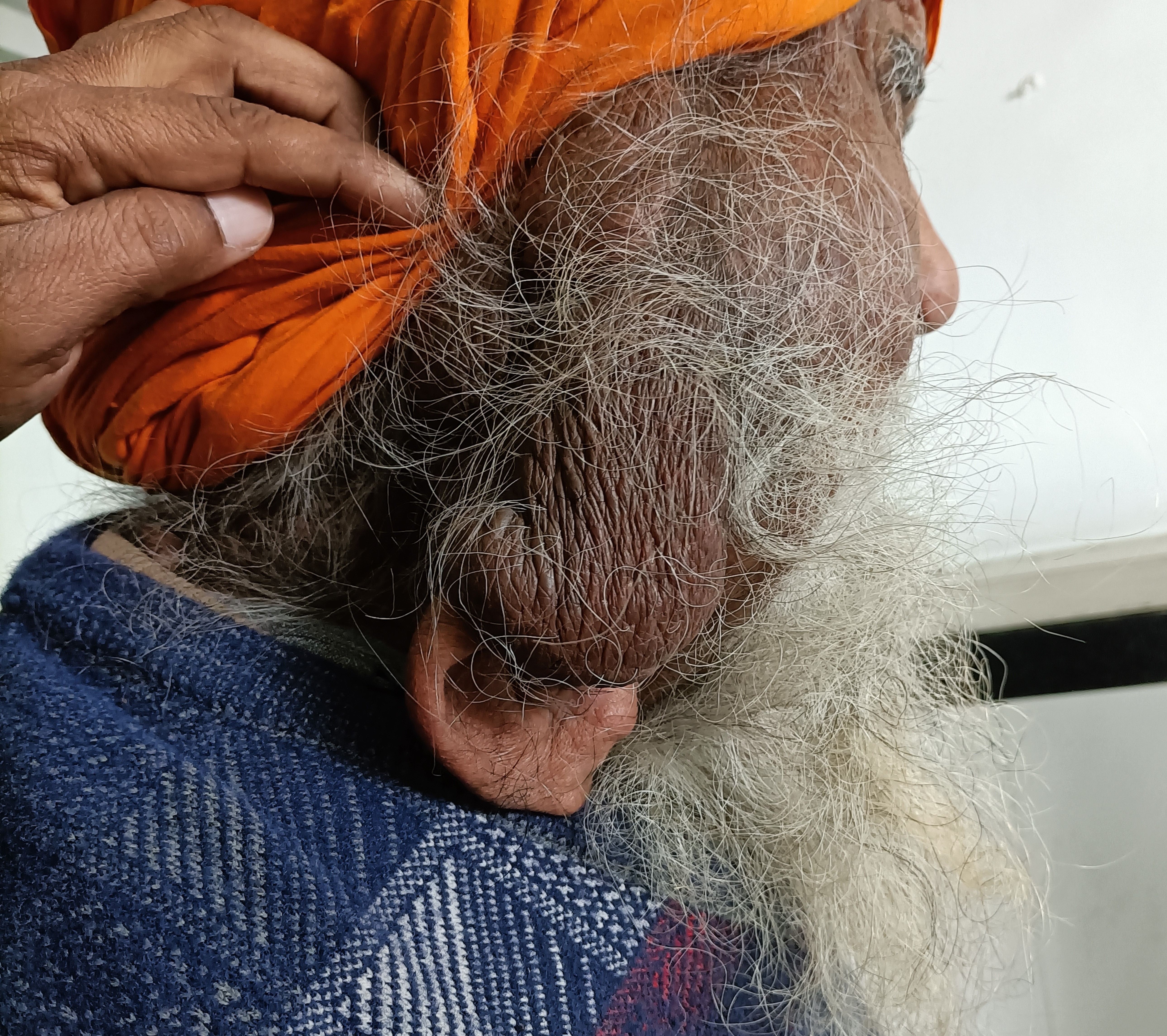

The facial lesion had brought down his pinna too, and he was unable to hear on that side. An MRI showed much deeper involvement, with destruction of the facial and cranial skeleton, suggesting malignant transformation. Was referred to neurosurgery department PGI.
Two bad experiences – messed up piles, fissure (Munchausen syndrome) and appendix cases. Examples of poor preoperative planning and preparation
Posted on: June 13, 2023
25.1.23
A 45 years old female had been posted for surgery for piles and a possible fissure too. At operation, the patient turned out to be very uncooperative, moving restlessly and talking incessantly and loudly. The anaesthetist had to use propofol to the spinal anesthesia to calm her down. An examination under anesthesia revealed no piles and no fissure, albeit a small perianal posterior skin tag was present. This was just excised. The anesthetist during the procedure had revealed her history of 4 previous surgeries for similar complaints. Munchausen syndrome was suggested as the possible diagnosis and was agreed to in the end with the treating physician.
The other case, a 45 years old male, was posted for appendicectomy. At operation, on palpation under anesthesia, a firm fixed mass was palpable. Again during surgery, the anesthetist gave the history! The patient had had pain for 4 months, was an opium addict and continued to neglect his symptoms. The incision had to be extended (RM), and a retrocecal chronic abscess and appendix were found with great difficulty. The pus was drained and the cavity washed clean after removing the appendix.
A 50 years old female was reportedly operated for cholecystectomy – an open operation, which was reportedly abandoned due to adhesions, as per the relatives’ version as there was no medical record available. However, a trial of laparoscopic approach was found to be quite easy and unremarkable. Possibly the operator was a beginner.
11.1.23
Another 50 years old lady had been having repeated scans (US and MRCP) reporting a contracted gallbladder with stones. The MRCP report also mentioned an ‘indentation’ on the main bile duct, suggesting pressure by a stone. At lap chole, the gallbladder was actually of normal size, albeit packed tightly with stones. The Calot’s dissection was difficult due to these stones, and the short cystic duct, dissected with difficulty, was ligated with no 1 vicryl.
27.12.22
A 50 years old male patient had to suffer for more than a year with different manifestations of gallstone disease:
Admitted on 21.11.22 by a local practitioner with acute pain RUQ of the abdomen, diagnosed as acute cholecystitis due to gallstones, largest stone being 14.5 mm. The ultrasound also reported wall thickening of the gallbladder wall, suggesting a longer history of the disease.

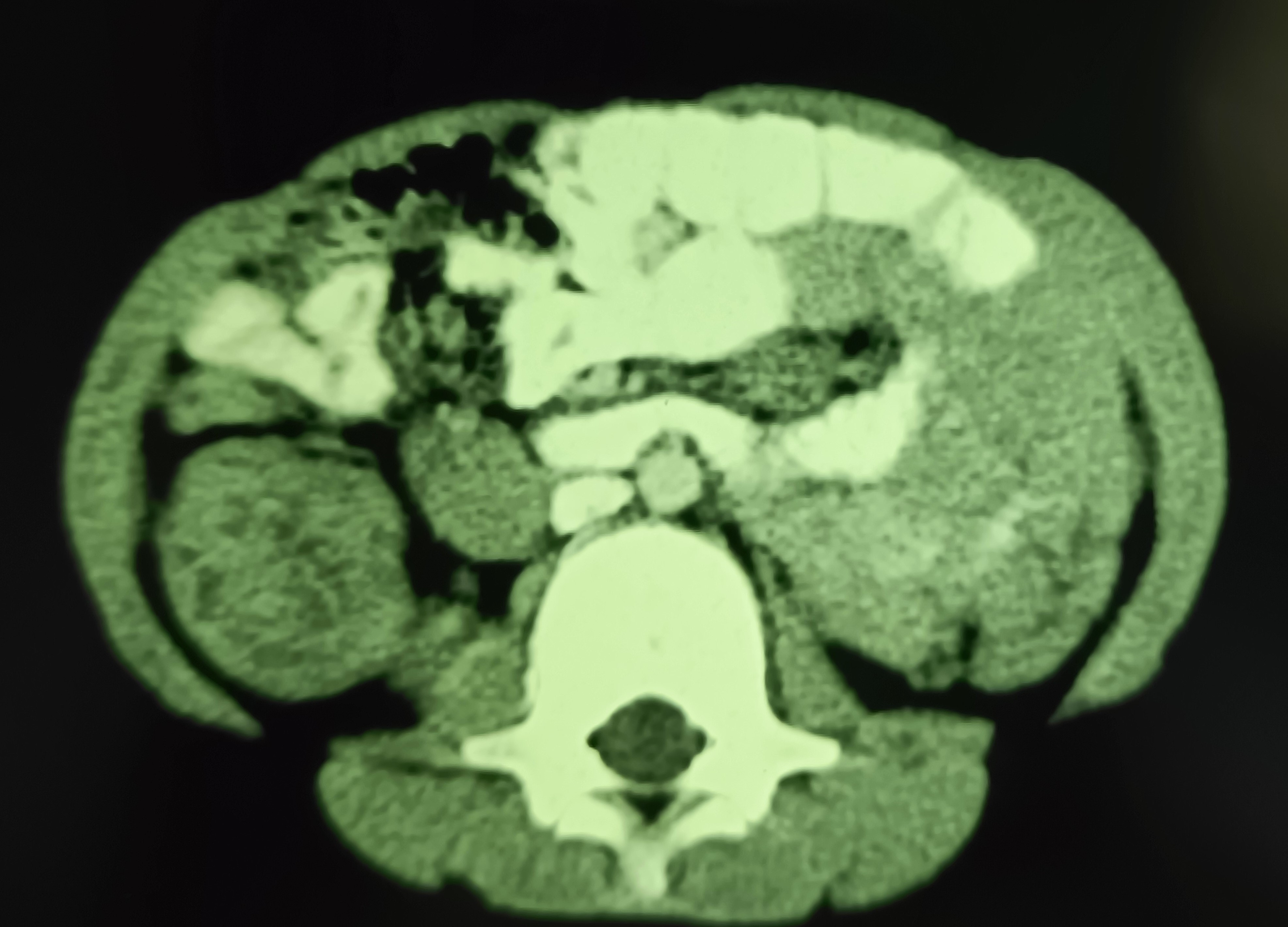
On 30.11.22, he was admitted again with a more severe attack of similar pain. This time, the ultrasound showed diffusedly thickened walls of the gallbladder, multiple stones and a possible collection in the fundal region, suggesting a perforation. A CT scan was suggested.

This reported distended gallbladder with diffusely thickened walls and multiple stones.
On 27.12.22 was admitted for lap chole. An USS this time showed a wall echo complex in the gallbladder fossa. A routine lap chole was performed, resulting in the end of the suffering.
