Piecemeal removal of thickwalled difficult gallbladders offers many advantages
Posted on: August 28, 2021
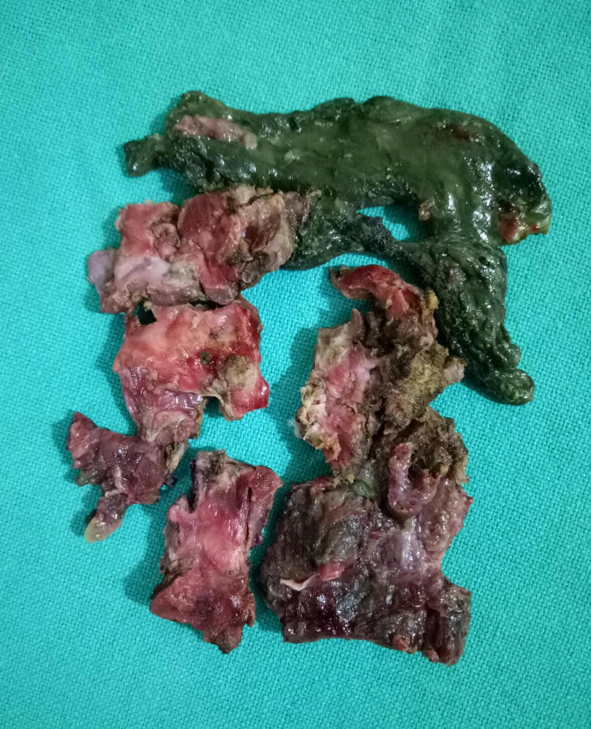
25.7.21
A thick-walled gallbladder (with empyema and some gangrene in the fundus) removed in several pieces through the epigastric port without dilating or enlarging the port;
this has been the practice now for many years with me as it offers so many advantages:
- Grasping the gallbladder which has grossly thick and tough walls and which is often very big and tense (sometimes empyema or mucocele) is impossible; so opening the fundus first of all helps in suctioning the mucus or pus and decreasing the size of the gall bladder. The large stones are also removed from within and the empty gall bladder is washed clean. Now the grasper can easily hold the left or the right flap.
- The dissection if easy in the Calot’s area can now be attempted and completed if it is easy. If not stop;
- Most often in these cases the infundibulum too is grossly thick and can’t be held with the grasper; so, the incision from the fundus is extended down to the infundibulum and the anatomy re-evaluated after the grasper can hold the fundus flap on the lateral or medial side.
- Since the gallbladder is now of manageable size and can be held comfortably and the posterior wall of the gallbladder viewed closely with the camera zoomed in, dissection can begin above the Rouviere’s sulcus level on the medial side high up near the middle of the body of the gallbladder with a hook separating the peritoneum if possible and then if easy, the Calot’s triangle can be dissected as usual. If not easy, again stop.
- More often than not, however, the peritoneum is fused with the thick walls of the gallbladder and no plane of dissection is safely available.
- In such cases, starting again high up, a plane can be created with a hook through the thickened walls of the gall bladder and then with a suction cannula and a piece of gauze as dissecting tools one can often go safely behind the posterior wall or behind the mucosa of the posterior wall to emerge on the lateral side.
- This plane of dissection (mostly submucosal, under the mucosa or through the thick wall) is then extended down similarly with gauze and hydrodissection, occasionally assisted by the hook to cut away a tough adhesion, always under clear and close vision of the camera, down to the infundibulum or the neck of gallbladder whichever appears to be the narrowest part which is then simply ligated with no 1 vicryl using extracorporeal knot. Before doing this one must again confirm through the opened up gallbladder that no stone is left behind in the gallbladder.
- The GB is simply divided just above the knot and removed, but the posterior wall of the gallbladder is left behind, the mucosa to be cauterized just before closure.
- The 2 big flaps of the gallbladder still be need to be divided into more vertical strips to make it easier to remove those strips through the epigastric port one by one; thus ensuring:
- The procedure remains minimal access still, and
- The troublesome problems of gas leak and the heightened risk of incisional hernia through the dilated/enlarged port are avoided.

Leave a comment